

 1 month ago
46
1 month ago
46
PROTECT YOUR DNA WITH QUANTUM TECHNOLOGY
Orgo-Life the new way to the future Advertising by AdpathwayWhen a 69-year-old man arrived at a hospital in Burkina Faso vomiting blood and complaining of diffuse abdominal pain, the clinical picture seemed straightforward. Upper gastrointestinal endoscopy revealed a large ulcer in the duodenal bulb, roughly 7 to 9 millimeters across and in active flare-up, and a stool test came back positive for Helicobacter pylori, the bacterium notorious for driving peptic ulcer disease. Physicians initiated standard eradication therapy, confident they were dealing with one of the most common diagnoses in gastroenterology. Yet the true cause of this patient’s hemorrhage lay elsewhere, hidden in the vast vascular network of the portal system, and only emerged days later when imaging revealed a rare and dramatic condition: a portal cavernoma, an intricate web of collateral veins that had formed in response to chronic blockage of the portal vein itself. The case, now published in the open-access journal Clinical Case Reports, offers a striking reminder that even seemingly obvious diagnoses can conceal a darker reality.
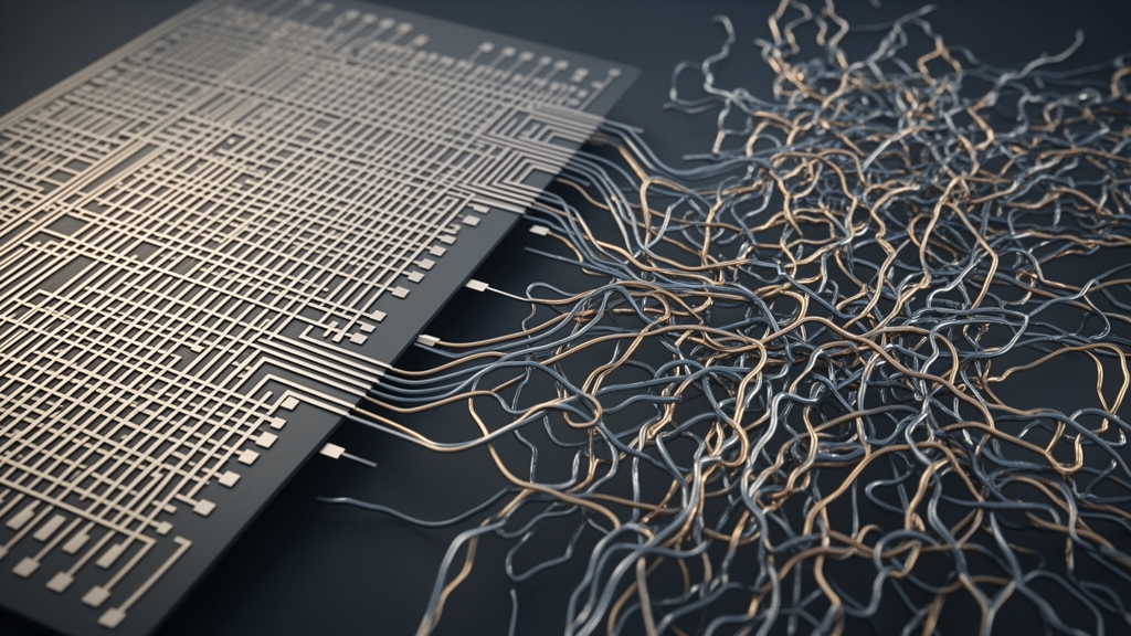
A portal cavernoma is defined as a network of veins, initially millimetric or even microscopic in size, that progressively dilates and through which hepatoportal blood continues to flow. It represents the body’s workaround for a problem: chronic occlusion of the extrahepatic portal system lasting more than three weeks. When the portal vein—the major vessel that channels nutrient-rich blood from the digestive organs to the liver—becomes irreversibly thrombosed, the surrounding tissue sprouts a tortuous meshwork of collateral channels to bypass the obstruction. First described in 1955 during the autopsy of a patient who died of mesenteric venous thrombosis, the condition has since become recognized as a significant cause of portal hypertension, but it remains sparsely documented in the African literature, particularly in sub-Saharan Africa, where only scattered case reports exist.
The patient’s initial presentation was deceptively ordinary. He had no particular medical history, yet he arrived with hematemesis—vomiting of blood—that had begun two days earlier, followed a day later by moderate diarrhea with blackish stools, a classic sign of digested blood passing through the gastrointestinal tract. There was no indication that he had consumed contaminated food or toxic substances. Clinical examination recorded a blood pressure of 100/60 mmHg, a heart rate elevated to 110 beats per minute, and a respiratory rate of 20 cycles per minute. His body mass index had fallen to 18 kg/m², signaling weight loss. Palpation revealed tenderness in the epigastric region, moderate ascites—fluid accumulating in the abdominal cavity—and an enlarged spleen consistent with Hackett stage II splenomegaly. Cardiac auscultation confirmed regular tachycardia at 110 beats per minute, a physiological compensation for volume loss.
Endoscopy performed shortly after admission revealed more than just the bulbar ulcer. The examination also identified grade IIa esophageal varices—dilated veins in the lining of the esophagus—without red signs, the endoscopic markers that predict imminent rupture. Additionally, the mucosa displayed a mosaic appearance, a hallmark of moderate portal hypertension. This combination should have raised a flag: peptic ulcers do not cause esophageal varices or mosaic mucosa, and both findings point toward elevated pressure in the portal venous system. Nevertheless, given the prominence of the ulcer and the positive H. pylori stool antigen test, the initial diagnosis of a bulbar ulcer was made, and eradication therapy was commenced. It was a reasonable start, but it addressed a contributor rather than the underlying engine of the disease.
The diagnostic turning point arrived two days later with an abdominal ultrasound. The study was consistent with heterogeneous portal vein thrombosis, accompanied by abundant ascites and, notably, a normal-sized, non-dysmorphic liver—an important observation, because cirrhosis is by far the most common cause of portal hypertension, and its absence demanded an alternative explanation. An abdominal computed tomography scan performed with portal vein injection then delivered the decisive findings. Contrast imaging revealed a heterogeneous filling defect in the portal trunk and its branches, with dilation of the vessels upstream of the obstruction, consistent with a thrombotic lesion of the portal trunk. The heterogeneous center of the lesion did not take up contrast, a pattern suggesting a portal cavernoma. The scan also demonstrated thrombosis of the splenic vein, a splenic nodule suggestive of an angioma, and an atrophic pancreas with a 9-millimeter dilation of the Wirsung duct, the main pancreatic duct.
Laboratory workup added nuance rather than contradiction. Transaminases were only slightly elevated, with AST at 44 IU/L and ALT at 48 IU/L, while the blood ionogram, complete blood count, and prothrombin level were all normal. Serology for HIV, hepatitis B, and hepatitis C came back negative, ruling out the viral infections that account for much of chronic liver disease worldwide. The etiological investigation, however, hit a practical wall: thrombophilia testing—including assays for protein C and S, antithrombin III, factor V Leiden, and the JAK2 mutation—could not be performed because these tests were simply unavailable in the clinical setting. This gap matters, because in adults the onset of portal vein thrombosis most often results from a combination of local and systemic prothrombotic factors, and guidelines call for systematic investigation of underlying thrombophilic disease.
With the imaging and clinical picture aligned, the team made their diagnosis: portal cavernoma revealed by gastrointestinal hemorrhage. The mechanism is elegant yet perilous. The portal obstruction creates a network of collateral veins of varying caliber through which hepatoportal blood is rerouted, but these fragile channels—and the varices they feed—operate under elevated pressure. In this patient, the hemorrhage was driven by rupture-prone esophageal varices, which occur in 90 to 95 percent of portal cavernoma cases, making variceal bleeding both the most common mode of presentation and the most feared complication. Other manifestations, including splenomegaly, abdominal pain, ascites, and transit disorders such as the patient’s diarrhea, are frequent but neither constant nor specific. The authors also point to portal hypertensive enteropathy and pancreatic atrophy as contributors to exocrine pancreatic insufficiency, which produces mucosal congestion, malabsorption, and diarrhea—a chain of physiological consequences that links seemingly unrelated symptoms back to the obstructed portal vein.
Treatment proceeded on several fronts simultaneously. The patient received 40 mg of injectable omeprazole every 12 hours to suppress acid and protect the ulcer, 40 mg of propranolol per day—a nonselective beta blocker that lowers portal pressure and provides primary or secondary prevention of variceal hemorrhage—and one sachet of Gaviscon every 8 hours. Endoscopic band ligation was performed to control the bleeding varices directly, and once hemorrhage was arrested, anticoagulation was initiated with 0.8 mL of enoxaparin every 12 hours, later transitioned to 4 mg of acenocoumarol daily with a target International Normalized Ratio of 2 to 3. Anticoagulation in portal cavernoma aims to prevent extension of thrombosis and, in some cases, to allow recanalization, though it must be balanced against the risk of rebleeding from varices.
The clinical evolution was favorable. Abdominal pain, ascites, and gastrointestinal bleeding all regressed, and the patient was discharged home after 10 days, following two stable INR readings of 2.3 and 2.5. Remarkably, no blood transfusion was required, as he remained hemodynamically stable with a hemoglobin level of 11 g/dL. He left the hospital on 40 mg of oral omeprazole twice daily and 4 mg of acenocoumarol, with anticoagulation planned for six months. At a three-month follow-up visit, he showed complete resolution of symptoms with a stable INR. Unfortunately, at the six-month mark the patient was lost to follow-up, and the team was unable to obtain follow-up imaging to assess whether the cavernoma had evolved, a sobering reminder of the practical challenges of longitudinal care in resource-limited settings.
The epidemiological context underscores just how unusual this case is. In the United States, the incidence of portal cavernoma is estimated at about 1 percent of the general population, based on large autopsy studies that documented portal vein thrombosis prevalence and lifetime risk across more than 23,000 consecutive examinations. In Africa, the true incidence is unknown but is thought to be below 1 percent according to the available literature. A Tunisian study in 2001 described just 19 observations over 25 years, while a Moroccan series reported 11 cases between 2003 and 2012. Most series show a male predominance, and the condition is overwhelmingly described in pediatric populations, where omphalitis—infection of the umbilical stump—remains a common cause of portal thrombosis in developing countries. In children, portal vein obstruction is also associated with growth retardation, attributed to reduced hepatoportal flow and resistance to growth hormone. Adult presentations in middle age, such as this one, are exceptionally rare in the published record.
The therapeutic hierarchy for portal cavernoma extends well beyond the medical management available to this patient. Beta blockers remain the cornerstone of hemorrhage prevention, but when endoscopic and pharmacological measures fail, ligation or sclerotherapy of varices is indicated, and in cases complicated by biliary symptoms—cavernomas can compress the bile ducts, producing a condition known as portal biliopathy—a porto-systemic shunt, with or without a porto-biliary shunt, may be required. Such surgical options demand infrastructure and expertise that are often unavailable in the settings where the disease is most likely to go undiagnosed. For the authors of this report, the case is a call to vigilance: when a patient presents with gastrointestinal hemorrhage and the endoscopy shows findings that exceed the usual scope of peptic ulcer disease, clinicians must look deeper into the portal vasculature. The ulcer, in this story, was real—but it was the supporting actor, not the star.
Subject of Research: A rare adult case of portal cavernoma caused by chronic portal vein thrombosis, revealed by gastrointestinal hemorrhage in a 69-year-old patient initially diagnosed with a bulbar ulcer.
Subject of Research: Medicine
Article Title: When a Bulbar Ulcer Hides a Darker Reality: The Unusual Diagnosis of a Portal Cavernoma in a 69-Year-Old Adult
Article References: Nacanabo, M. W., Seghda, A. A. T., Yaméogo, A. A., Lengani, E. H., Zingué Ouattara, A. B., Guiarra, A., Bayala, Y. L. T., Porgo, A. N., Aziz, A., Traoré, A. D., Tall/Thiam, A., Millogo, G. C., Yaméogo, V. N., & Samadoulougou, A. K. (2026). When a Bulbar Ulcer Hides a Darker Reality: The Unusual Diagnosis of a Portal Cavernoma in a 69‐Year‐Old Adult. Clinical Case Reports, 14(7), Article e73040. https://doi.org/10.1002/ccr3.73040
Image Credits: AI Generated
DOI: 10.1002/ccr3.73040
Keywords: portal cavernoma, portal vein thrombosis, portal hypertension, esophageal varices, gastrointestinal hemorrhage, Helicobacter pylori, bulbar ulcer, adult diagnosis, anticoagulation, sub-Saharan Africa, Clinical Case Reports
Cite Scienmag News
APA MLA Chicago
Ophelia Keating. (September 4, 2026). Bulbar Ulcer Reveals Rare Portal Cavernoma Diagnosis in 69-Year-Old Patient. Scienmag. https://scienmag.com/bulbar-ulcer-reveals-rare-portal-cavernoma-diagnosis-in-69-year-old-patient/
Copy citation Download RIS
Tags: cavernous transformation of the portal veinchronic portal vein occlusionclinical case of portal cavernomaclinical case reportduodenal ulcerduodenal ulcer in elderlyendoscopic findings in ulcer diseasegastric ulcer bleedinggastrointestinal bleedinggastrointestinal hemorrhage causesgastrointestinal hemorrhage diagnosisHelicobacter pylori in ulcerHelicobacter pylori infectionhepatic portal system anomaliesPortal cavernomaportal cavernoma diagnosisportal veinportal vein thrombosisrare causes of upper GI bleedingrare vascular conditionsvascular collateral formationvascular complications of portal vein blockage



















 English (US) ·
  French (CA) ·