
 9 hours ago
9
9 hours ago
9
PROTECT YOUR DNA WITH QUANTUM TECHNOLOGY
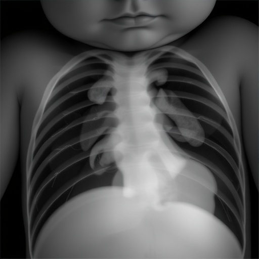
Orgo-Life the new way to the future Advertising by AdpathwayIn an illuminating breakthrough published recently in Pediatric Research, scientists have unveiled the intricate cardiac adaptations that occur in fetuses diagnosed with congenital diaphragmatic hernia (CDH). This developmental anomaly, characterized by an incomplete formation of the diaphragm, leads to significant pulmonary hypoplasia and resultant cardiovascular stress, posing a daunting challenge for neonatal survival. The research, led by Bhattacharya and Patel, offers an unprecedented glimpse into the mechanobiology of fetal heart remodeling under abnormal pressure conditions, deepening our understanding of the pathophysiology underlying CDH.
Congenital diaphragmatic hernia disrupts normal intrathoracic anatomy, allowing abdominal organs to encroach upon the thoracic space. This intrusion severely compromises lung development, inducing hypoxic stress and hemodynamic alterations that drastically elevate pulmonary vascular resistance. The fetal heart, especially the right ventricle, is subjected to augmented afterload, prompting compensatory structural and functional remodeling. Researchers have long grappled with decoding these adaptive processes, which are pivotal determinants of postnatal survival and long-term cardiopulmonary outcomes.
Bhattacharya and Patel harnessed state-of-the-art imaging and molecular techniques to dissect the cardiac adaptations occurring in utero among CDH cases. High-resolution fetal echocardiography combined with innovative computational modeling allowed them to quantify changes in ventricular geometry, wall stress, and myocardial strain patterns. Their findings reveal that the right ventricle, confronted with increased pressure, undergoes hypertrophic remodeling characterized by augmented myocardial thickness and altered fiber orientation. This structural adaptation ostensibly aims to preserve cardiac output in the face of escalating pulmonary vascular resistance.
Interestingly, the researchers also observed a concomitant reduction in right ventricular compliance, indicating increased myocardial stiffness. This biomechanical shift is hypothesized to be a double-edged sword; while augmenting contractile strength, it may eventually predispose the heart to diastolic dysfunction. The delicate balance between adaptive hypertrophy and maladaptive fibrosis emerges as a critical juncture in fetal cardiac pathophysiology within CDH, underscoring the need for refined therapeutic interventions that can modulate this process.
Further molecular analyses pinpointed upregulation of mechanosensitive signaling pathways, including those mediated by transforming growth factor-beta (TGF-β) and angiotensin II, which orchestrate fibrotic remodeling and extracellular matrix deposition. The fetal myocardium’s response to escalating pressure involves a complex dialogue of pro-hypertrophic and pro-fibrotic cues, which ultimately sculpt the shape and function of the developing heart. These signaling networks represent promising therapeutic targets for mitigating adverse cardiac remodeling in CDH.
The study also addresses the temporal dimension of cardiac adaptation. The authors demonstrate that myocardial remodeling initiates early in gestation and progresses in tandem with the severity of pulmonary hypoplasia. This temporal correlation underscores the potential for early in utero interventions designed to alleviate pressure overload or modulate maladaptive signaling cascades, thereby improving cardiac resilience and neonatal outcomes.
Notably, the research highlights the heterogeneity of cardiac responses within the CDH population. Variability in ventricular remodeling patterns appears to correlate with differences in clinical severity and survival rates, suggesting that individualized assessments of cardiac mechanics could inform prognosis and guide perinatal care strategies. This paradigm shift towards precision medicine in fetal cardiology has far-reaching implications for optimizing the timing and nature of interventions.
Bhattacharya and Patel’s investigation further reveals that left ventricular morphology is not spared; secondary adaptations occur due to altered loading conditions and interventricular interactions. The left ventricle may exhibit subtle geometric deformation and impaired compliance, reflective of the complex hemodynamic interplay within the compromised thoracic environment. These cross-ventricular effects deepen our understanding of global cardiac remodeling in CDH.
The comprehensive approach integrates advanced imaging with molecular biology, offering a multidimensional view of fetal cardiac adaptation. Such integrative methodologies are at the frontier of developmental cardiology research, allowing for the translation of mechanistic insights into clinical practice. This synergy between bench and bedside opens new vistas for fetal therapy in CDH.
Clinicians and researchers alike stand to benefit from these insights, which pave the way for novel interventions. Potential strategies might include pharmacologic modulation of fibrosis pathways, fetal surgical techniques to decompress the thorax, or innovative biologic agents targeting myocardial signaling. The ultimate goal is to normalize intrathoracic pressures and optimize cardiac development before birth.
Moreover, this research has significant implications for postnatal management. Understanding the prenatal trajectory of cardiac remodeling can help anticipate complications such as pulmonary hypertension and right heart failure after delivery. It also informs long-term surveillance and therapeutic planning for survivors of CDH, who often face chronic cardiopulmonary challenges.
The findings presented in this landmark study underscore the critical importance of multidisciplinary collaboration, combining fetal medicine, cardiology, imaging science, and molecular biology. Such collaboration is essential to unravel the complexities of congenital anomalies and develop effective treatments that improve quality of life from the earliest stages of development.
In conclusion, the work by Bhattacharya and Patel represents a seminal advance in our grasp of how the fetal heart contends with the mechanical burden imposed by congenital diaphragmatic hernia. By elucidating the structural and molecular underpinnings of cardiac adaptation under pressure, this research not only enhances fundamental knowledge but also inspires hope for innovative therapies that can rewrite the destiny of affected infants.
Subject of Research: Cardiac adaptation and remodeling in fetuses diagnosed with congenital diaphragmatic hernia under altered hemodynamic pressure conditions.
Article Title: Born under pressure: cardiac adaptation in congenital diaphragmatic hernia.
Article References:
Bhattacharya, A., Patel, N. Born under pressure: cardiac adaptation in congenital diaphragmatic hernia. Pediatr Res (2026). https://doi.org/10.1038/s41390-026-05257-0
Image Credits: AI Generated
DOI: https://doi.org/10.1038/s41390-026-05257-0
Tags: cardiopulmonary outcomes congenital diaphragmatic herniacomputational modeling cardiac functioncongenital diaphragmatic hernia cardiac adaptationfetal echocardiography in congenital defectsfetal heart remodeling in CDHintrathoracic anatomy disruption CDHmechanobiology of fetal heartmyocardial strain in fetal developmentneonatal survival congenital anomaliespulmonary hypoplasia cardiovascular impactpulmonary vascular resistance fetal heartright ventricle compensatory remodeling


 English (US) ·
  French (CA) ·