
 15 hours ago
4
15 hours ago
4
PROTECT YOUR DNA WITH QUANTUM TECHNOLOGY
Orgo-Life the new way to the future Advertising by Adpathway
Published on August 1st, 2025 | by David Marshall
Determining the origin of teeth in vertebrates is an incredibly significant but notoriously difficult problem within palaeontology. Teeth didn’t evolve in the mouths of our ancestors, but are first seen as part of the external skeletons of jawless fish as structures called ‘odontodes’. These would later migrate into the mouth with the evolution of jaws, becoming the teeth we have today, but odontodes still remain present in the skin of modern cartilaginous fish, giving them their rough texture.
The oldest known odontodes are from the late Cambrian Period and represent the very first evidence for vertebrates in the fossil record. Unfortunately, they are only ever found as part of fragmentary pieces of exoskeleton, however, given that their specific construction is only known in vertebrates, there is little else they could possibly be…
Joining us for this episode is Dr Yara Haridy, University of Chicago, who set out to use modern new scanning techniques to better understand the nature of these first teeth and what they tell us about the evolution of vertebrates. What she discovered was unexpected, but also led to better understanding of the purpose of odontodes in the dermal exoskeletons of our ancestors. Her research was recently published in Nature and is free to access.
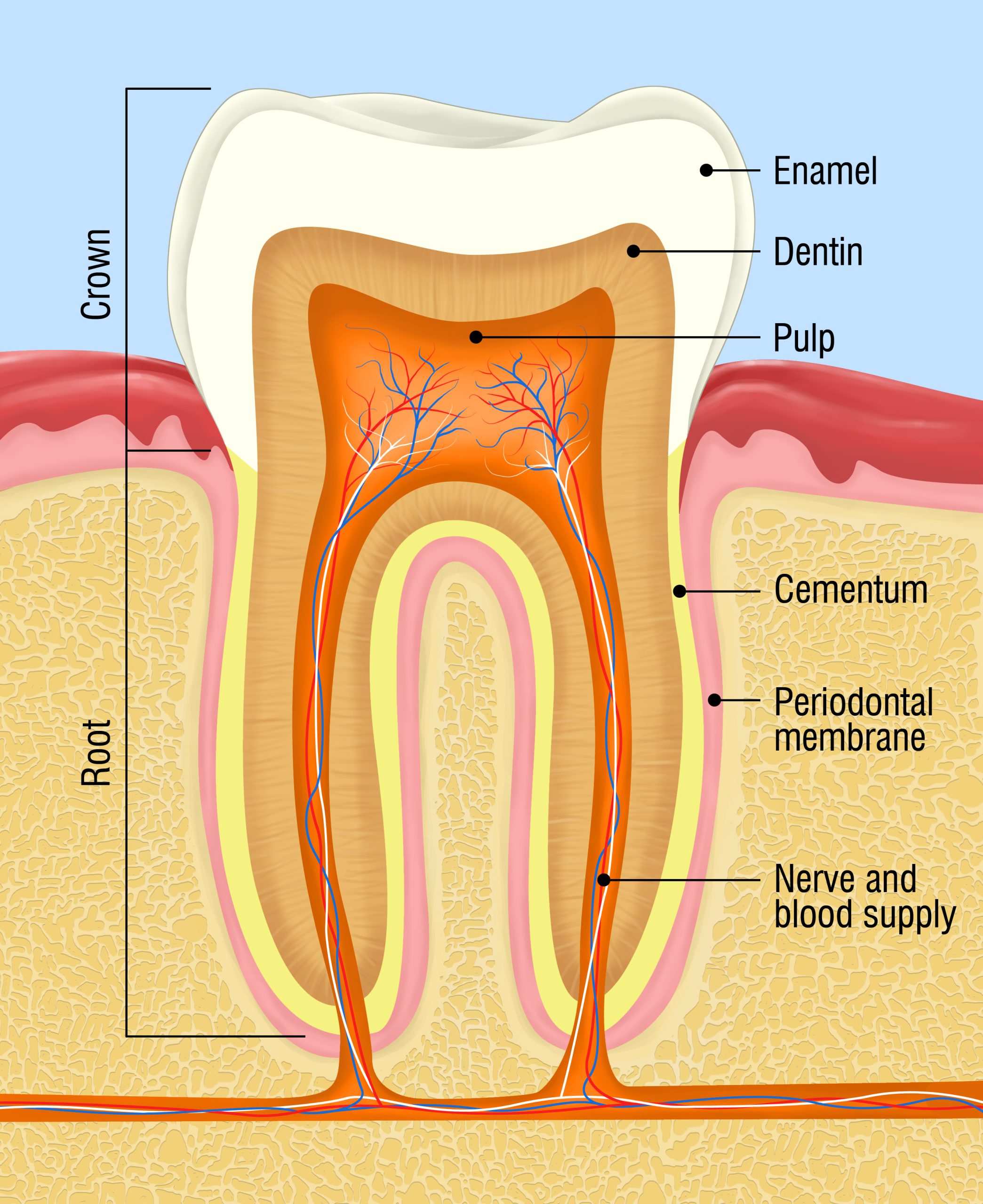 Our teeth are complex structures made out of enamel and dentine [dentin, US spelling] surrounding a pulp cavity. They are innervated so that they are sensitive to different stimuli and are in possession of their own blood supply. Enamel and dentine are only known in vertebrates and since they are some of the hardest biological materials known, teeth have a significantly better chance of being fossilised than other softer tissues. Teeth therefore play a critical role in our understanding of vertebrate evolution.
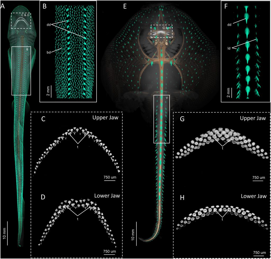 Teeth are not just restricted to the mouth, but are also found within the skin of chondrichthyans (cartilaginous fish e.g. sharks and rays) as denticles/scales. Whether they are in the mouth or in the dermal skeleton, they all share the same gene regulatory network and so are ‘homologous’ (of the same evolutionary origin, even if later used for different purposes) and are collectively referred to as ‘odontodes’.
Image: Micro-computed tomography (μCT) scans highlighting the diversity odontodes in elasmobranch embryos: A-D small-spotted catshark (Scyliorhinus canicula) and E-H Little skate (Leucoraja erinacea). Credit: Nicklin, et. al. 2024. Evolution, development, and regeneration of tooth-like epithelial appendages in sharks. Developmental biology, 516, pp.221-236. CC BY-NC 4.0.
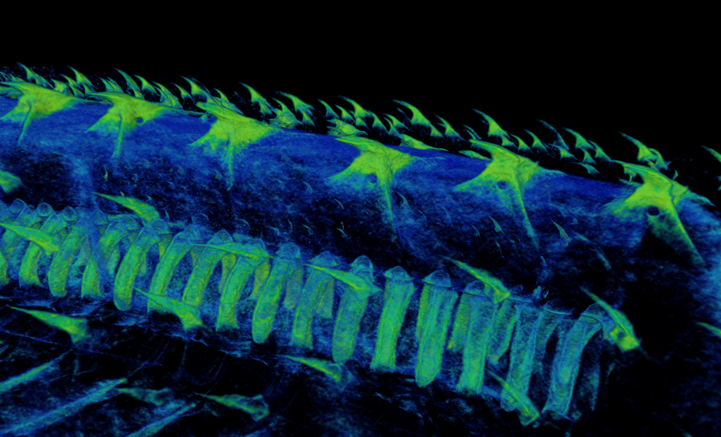 Odontodes in the dermal skeleton have a different function to those in the mouth and are used for protection, hydrodynamics and likely sensory purposes.
 Image: CT scan image of tooth like dermal denticles on a catshark. Credit: Dr Yara Haridy /University of Chicago.
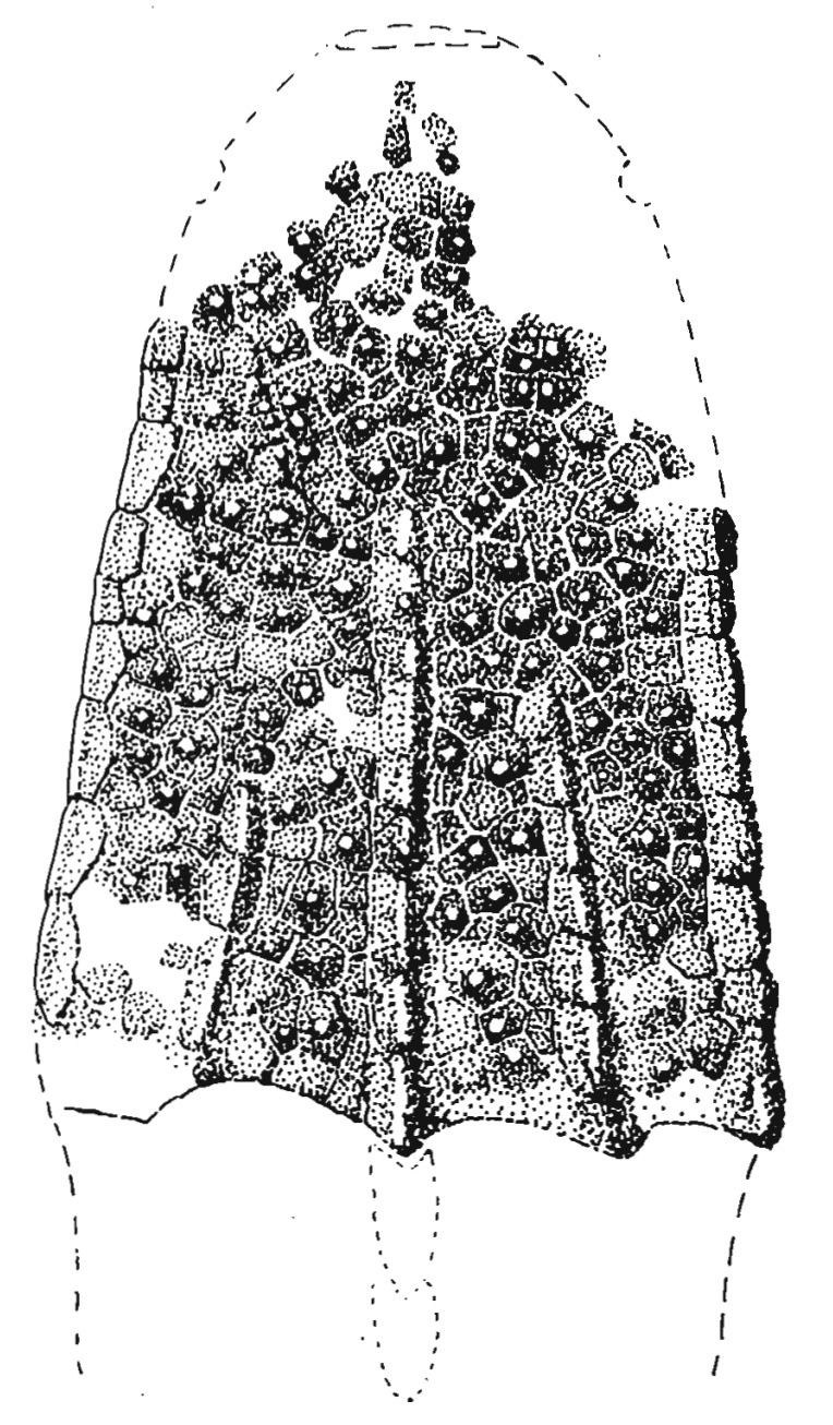 Odontodes are known from the external skeletons of jawless fish and are found well before the evolution of jaws and the first oral odontodes. The bony plates on the headshield of Astraspis desiderata were capped with an enameloid, thus showing this material first evolved externally.
 Credit: Tarlo, L.B., 1962. The classification and evolution of the Heterostraci. Acta Palaeontologica Polonica, 7(1-2). CC BY 4.0.
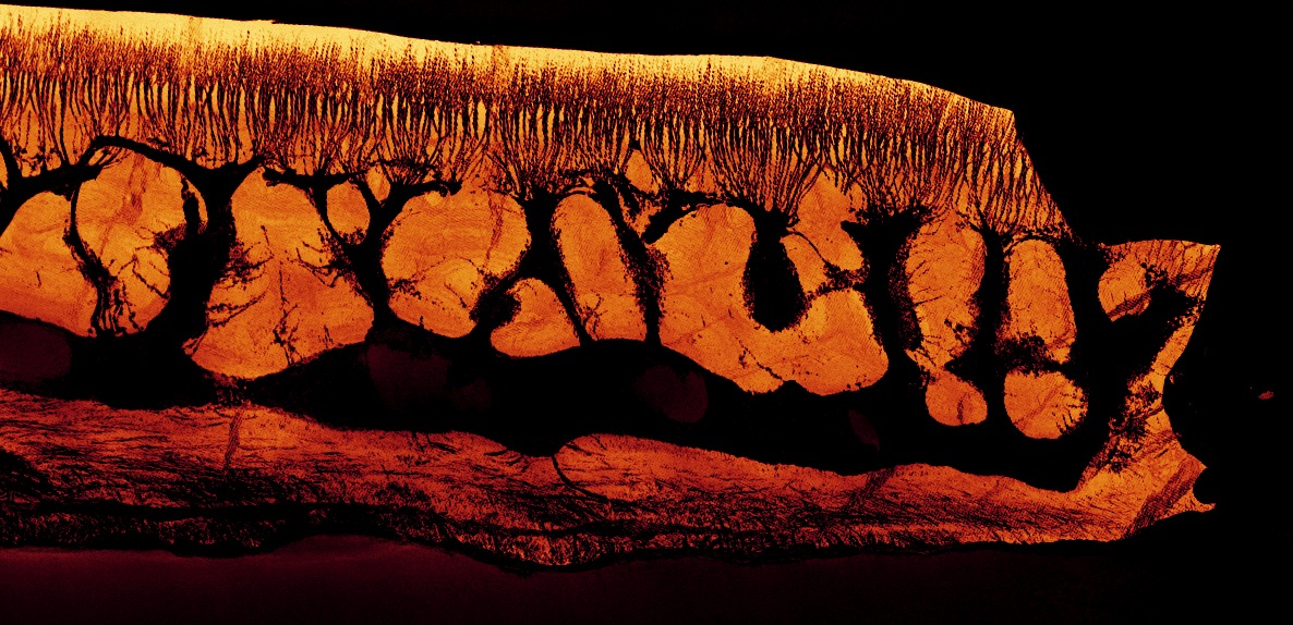 A virtual thin section from a synchrotron scan of an odontode from the jawless fish Lepidaspis. The larger dark areas within the odontode are pulp cavities, whereas the thinner branches are ‘dentine tubercles’ where a dentine-secreting cell would have reached up to secrete dentine. The colour of the image reflects the density of material, with lighter colours being higher densities. This shows the density dentine towards the upper surface. Credit: Joe Keating, University of Bristol.
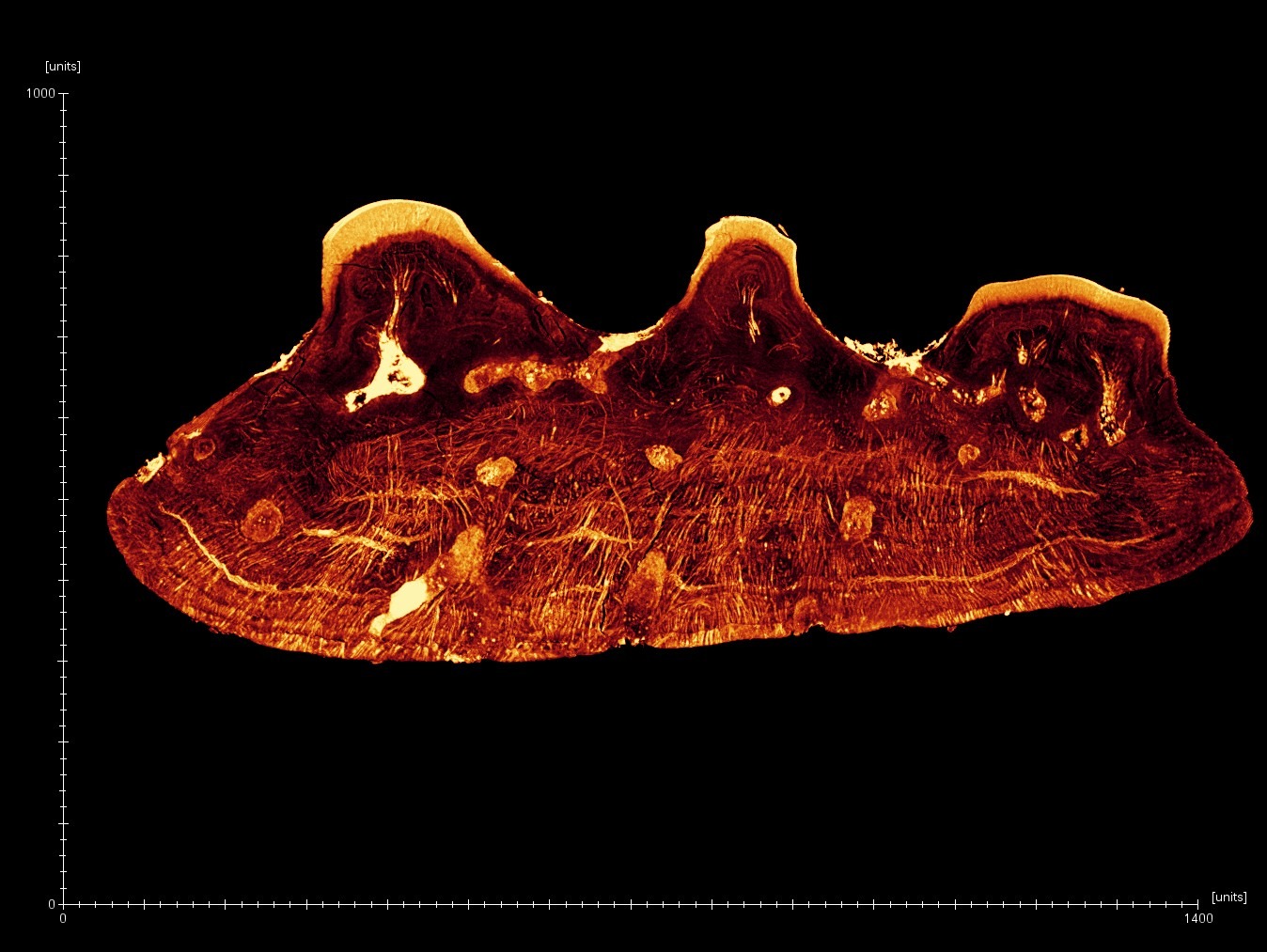 Odontode from Tesseraspis clearly showing bright enameloid caps on the top surface. The texture in the lower half is caused by the fibres that anchor the odontode within the skin of the fish. Credit: Joe Keating, University of Bristol.
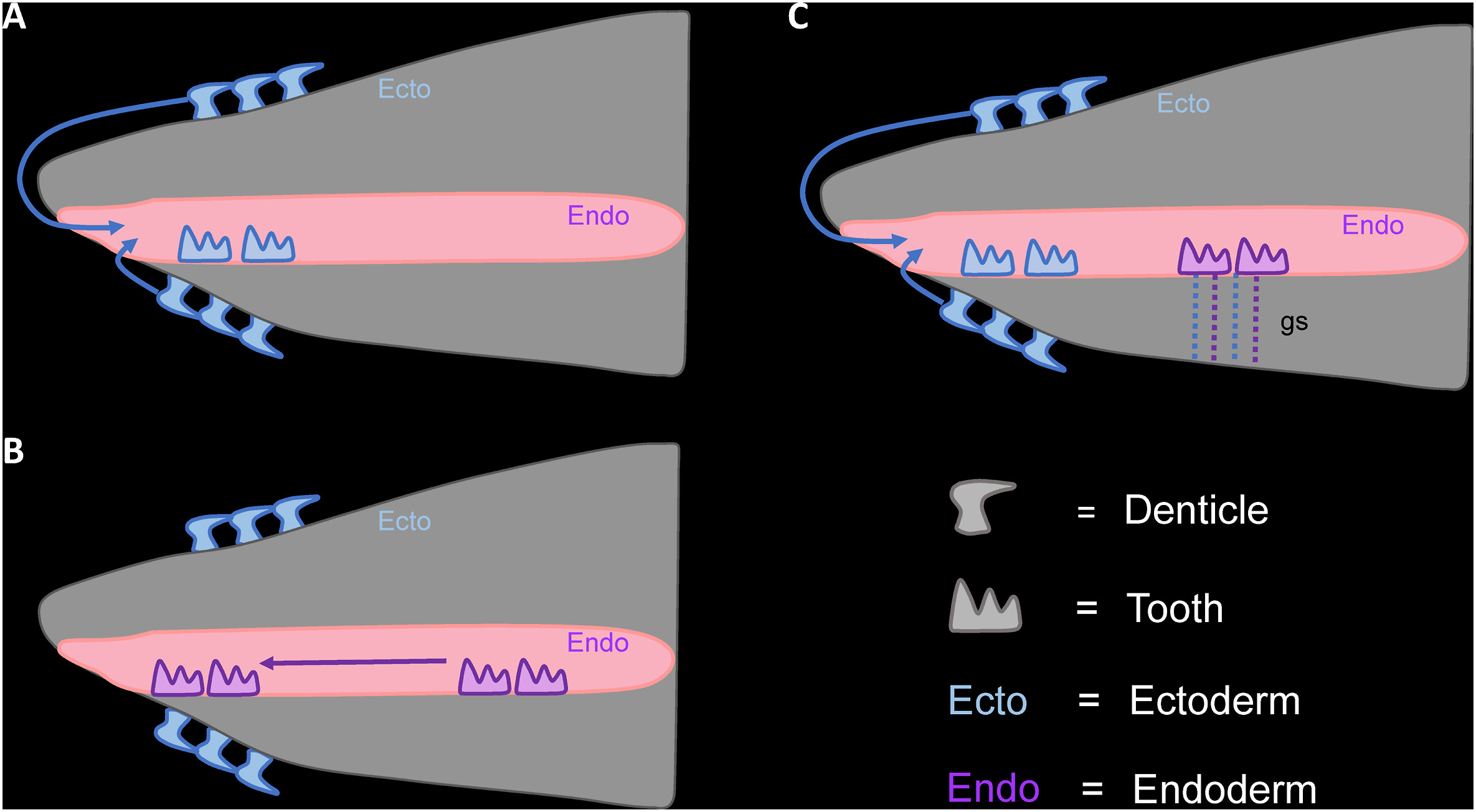 Since we know all odontodes are all homologous, there have been different theories developed to explain why we see them first externally and how they might have been incorporated internally. A, Outside-in theory – odontodes develop externally and migrate into the mouth and pharynx (throat); B, Inside-out theory – odontodes develop in the pharynx and migrate into the mouth, whilst external odontodes are derived independently; C, Modified outside-in theory – odontodes are (additionally) internalised through the gill slits (gs).
Credit: Nicklin, et. al. 2024. Evolution, development, and regeneration of tooth-like epithelial appendages in sharks. Developmental biology, 516, pp.221-236. CC BY-NC 4.0.
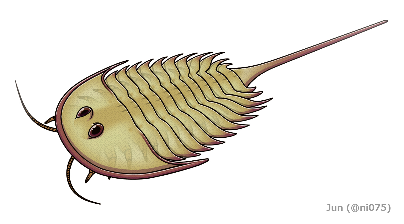 The oldest evidence for vertebrates in the fossil record comes from the late Cambrian and is identified from fragments of exoskeleton bearing odontodes. These fragments were thought to be from a jawless fish which was given the name Anatolepis. This claim has been contested by some researchers who suggested that this material might actually be from an aglaspidid arthropod. Aglaspidids are artiopods and so are closely related to trilobites and are interpreted to have had a similar way of life. Unlike trilobites, whose exoskeletons are composed of calcium carbonate, aglaspidids use calcium phosphate: the same material vertebrate bone is made of. Additionally, the exoskeleton possesses raised bumps called ‘tubercles’ which are of a similar shape and size to the odontodes of Anatolepis. Image: Reconstruction of Aglaspis spinifer. Credit: Jun CC BY-SA 4.0.
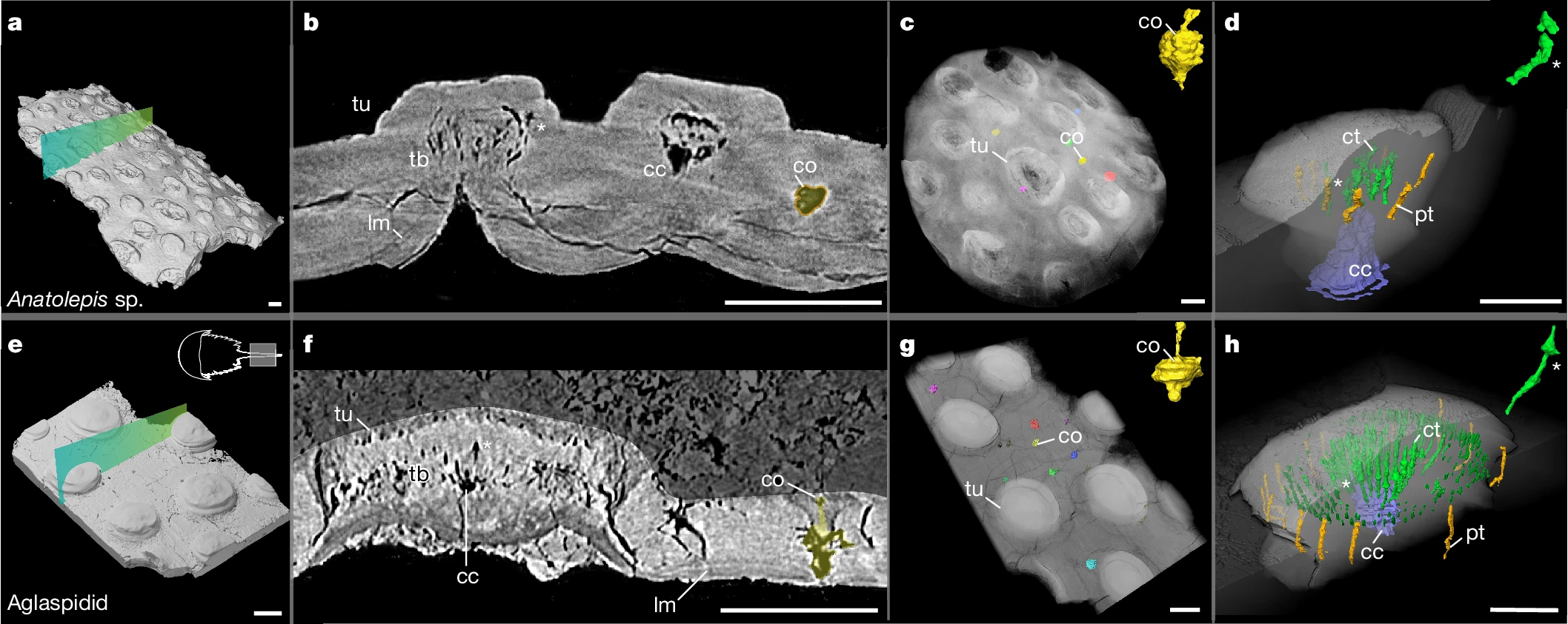 Through synchrotron micro-CT scanning, Dr Haridy was able to create high resolution 3D images of the different fossil material for comparison. The results show so little difference between Anatolepis (top row) and an aglaspidid (bottom row) that it can be shown that ‘Anatolepis’ is actually just fragmentary remains of aglaspidid exoskeleton and what were thought to be odontodes were in fact tubercles. These two structures have very different evolutionary origins but have a similar appearance/function and this is what we term ‘convergence’ (which is essentially the opposite of homology).
Key: cc, central cavity (purple); co, cuticular organ (yellow); ct, central tubules (green); lm, lamellar tissue; pt, peripheral tubule system (orange); tb, tubules; tu, tubercle. Scale bars, 100 μm. Credit: Haridy et. al. 2025. The origin of vertebrate teeth and evolution of sensory exoskeletons. Nature, pp.1-6. CC BY-NC-ND 4.0
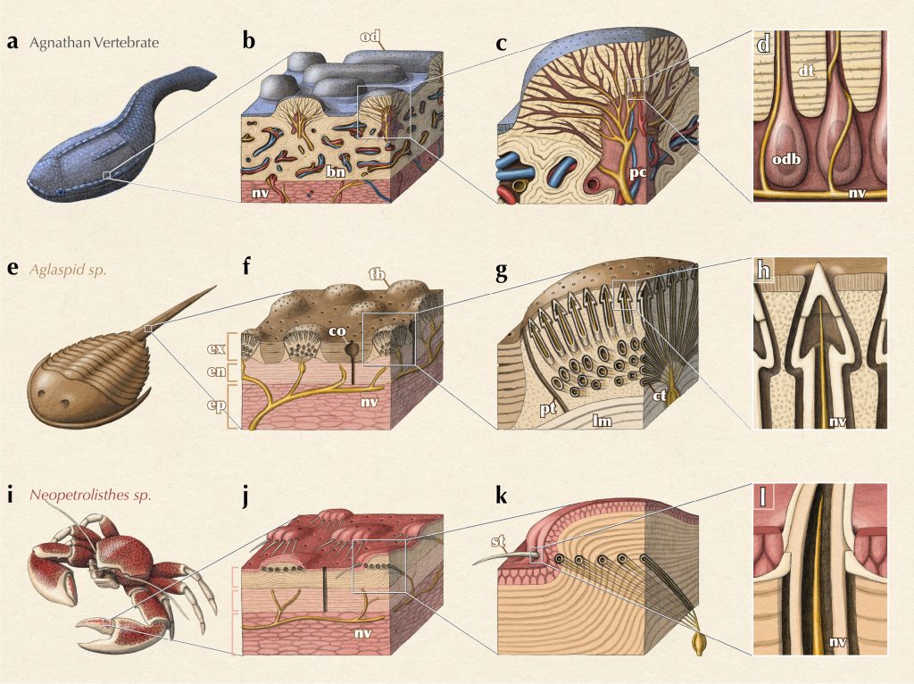 The exoskeletons of the different groups can be summarised as above. In modern arthropods, such as the porcelain crab Neopetrolisthes, tubercles are known to have a sensory function since they are innervated and will often house sensory hairs called setae. Given the similarity of their exoskeletons and tubercles, It is likely that the aglaspidids had a similar sensory function for their tubercles. So if form follows function, could it be the case that vertebrate odontodes also served a sensory function in a remarkable case of convergent evolution? Credit: Alex Boersma/ https://www.alexboersma.com.
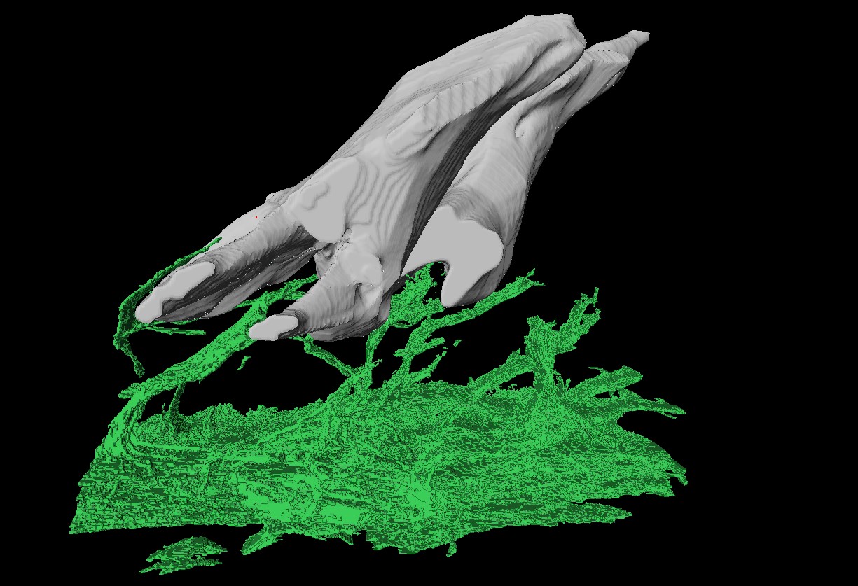 Modern chondrichthyan odontodes were imaged and were demonstrated to be innervated.
 Image: Segmented confocal scan of the tooth-like-odontode structure from suckermouth catfish fish, showing nerves (in green) that allow transmission of sensory information from the tooth like odontode to the nervous system. Credit: Dr Yara Haridy /University of Chicago.
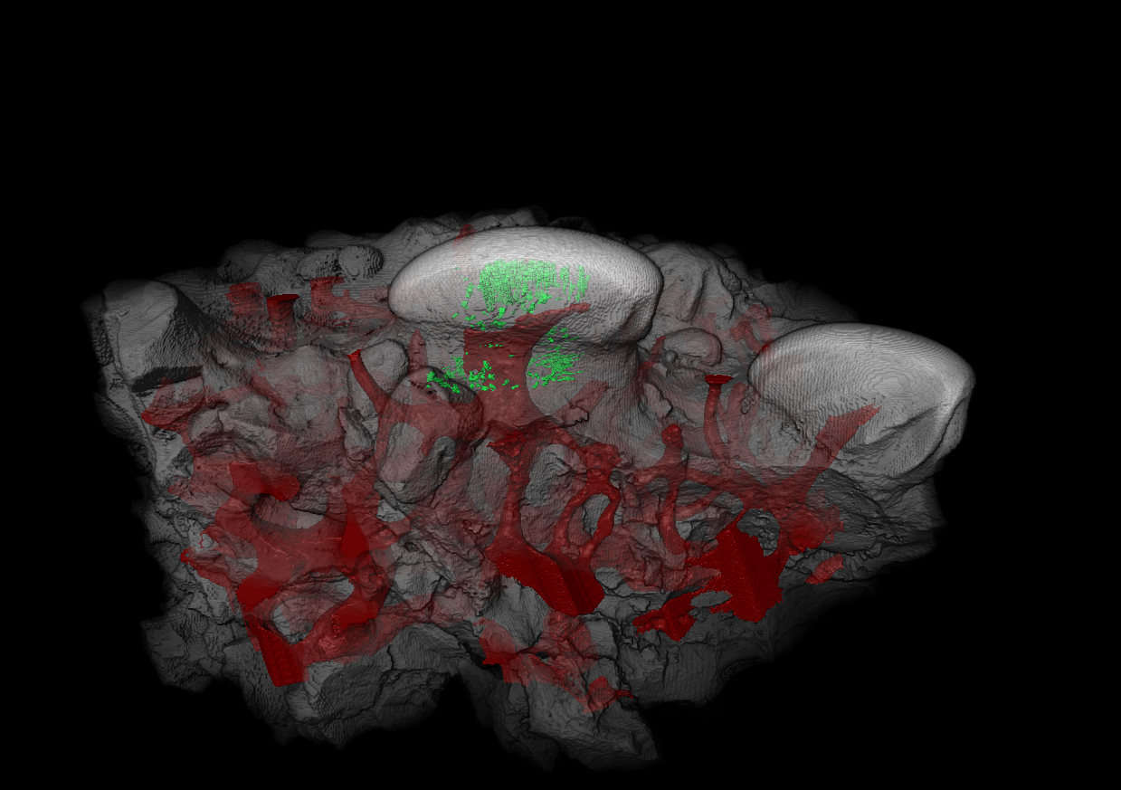 Subsequent scans of Astraspis odontodes showed a similar innervation and vasculature (red), thus confirming that when odontodes first appeared, they were able to serve a sensory function like modern teeth. Image: CT scan of the tooth-like-odontode structure from Astrapsis. The tubules (green) are filled with dentine, the same material that makes up the sensitive inner layer of modern teeth. In red is the vascular system which would have housed the nerves in life allowing for sensation to be transmitted. Credit: Dr Yara Haridy /University of Chicago.
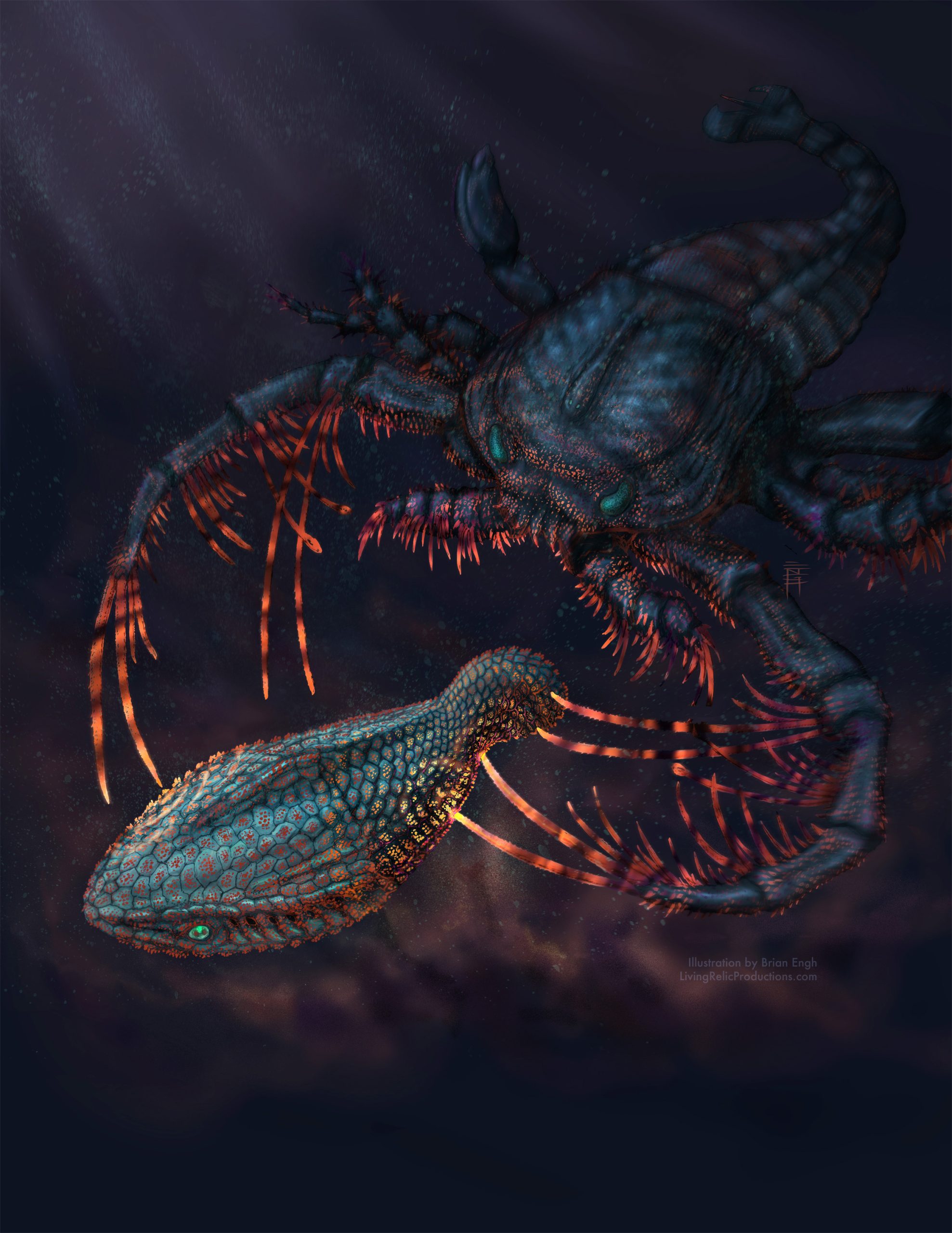 Artistic rendering of the sensory exoskeletons of the early jawless vertebrate Astraspis being attacked by the sea-scorpion Megalograptus in dark shallow waters. The pulsating glow of the interacting exoskeletons representing the convergent ability of sensation. Credit: Brian Engh/ https://www.livingrelicproductions.com.
Tags: Aglaspidid, agnathan, convergence, CT, exoskeleton, gnathostome, homology, jawless fish, odontodes, sensory ecology, synchrotron, Teeth, Vertebrates


 English (US) ·
  French (CA) ·