

PROTECT YOUR DNA WITH QUANTUM TECHNOLOGY
Orgo-Life the new way to the future Advertising by Adpathway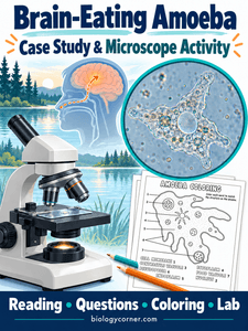
A summer swim turns into a medical mystery in this activity about amoebas. Students begin with a short case study about Naegleria fowleri, often called the “brain-eating amoeba.” Then they examine a preserved amoeba under the microscope. The lesson connects a real event with cell structures students can observe and additional practice with the microscope.
The case study follows the documented death of an eight-year-old boy after swimming in Nebraska’s Elkhorn River in 2022. Students learn how contaminated water entering the nose can allow the amoeba to reach the brain, where it causes tissue damage and swelling. Reading questions help students identify key details, explain how infection occurs, and connect environmental conditions with human health. The case also offers an opportunity to discuss the difference between a disease being rare and being deadly.
For the microscope portion, students view a prepared slide of preserved Chaos carolinensis, or giant amoeba. This is a different species from the organism described in the case study. It can easily be obtained for biological supply companies. Students practice locating a specimen at low power, increasing magnification, and drawing what they observe. They look for the cell boundary, cytoplasm, pseudopodia, and internal structures such as food vacuoles.
Overview of the Activity
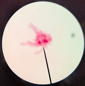
I included this activity with the chapter on “Humans and Global Change” by Miller and Levine (Bee Book). This adds a little bit of a break to classroom lectures and gives students additional practice with the microscope. They can also tie the case to human impacts and changes in climate. Though you could put it anywhere students might just need a little hands-on experience.
The activity focuses on structure rather than movement, because the specimen is preserved. You can order live amoeba, but in my experience, they don’t last long and students can have difficulty finding them. My goal with this activity is to give students more practice with the microscope while also connecting it to real-world events.
An amoeba coloring page reinforces vocabulary, including the cell membrane, food vacuole, and pseudopodia. Students connect these structures with their functions, such as capturing food and removing excess water. My students will explore the cell after the unit on ecology, so for this section, I help them label the amoeba. Though the coloring has each structure numbered, and students only need to find it from the list and color it to match.
Use the resource during a unit on protists, cells, or microscope skills. The reading and coloring page can introduce the topic before the lab, or students can complete them while rotating through microscope stations. Together, the activities give students a chance to read, observe, draw, and connect biology to a real-world event.
Related Resources
Amoeba Coloring – this image is included in the lab activity, but you can also use it as a stand-alone assignment
Amoeba and Paramecium Virtual Lab – students view the movements of two protists from a video and compare
Basic Microscope Lab – examine the letter E to learn how to use the microscope
Giant Microbes Amoeba – this fun plush can help students visualize the organism
















 English (US) ·
  French (CA) ·