

 2 hours ago
11
2 hours ago
11
PROTECT YOUR DNA WITH QUANTUM TECHNOLOGY
Orgo-Life the new way to the future Advertising by AdpathwayDamaged mitochondria may be the hidden hand that turns slow cellular wear into the chronic inflammation of old age. A wide-ranging review published in GeroScience by Salvatore Nesci of the University of Bologna and Cristina Algieri of Link University in Rome lays out a detailed mechanistic argument that failing mitochondria are not merely casualties of ageing but active organisers of it, coupling bioenergetic collapse to cellular senescence and to the low-grade, sterile inflammation that researchers call inflammaging. The authors assemble evidence from respiratory biochemistry, mitochondrial genetics, cell death research and immunology into a single feed-forward model in which the same organelle that powers the cell becomes, when it breaks down, the spark that keeps inflammatory signalling smouldering across tissues.
The starting point of the review is familiar but consequential: as the electron transport chain loses efficiency with age, proton pumping across the inner mitochondrial membrane falters, the membrane potential falls and ATP output declines. At the same time, electrons leak preferentially at respiratory complexes I and III, feeding the production of reactive oxygen species. These oxidants then attack mitochondrial DNA, respiratory-chain proteins and membrane lipids, further degrading oxidative phosphorylation and locking the organelle into a self-amplifying spiral of bioenergetic failure and oxidative stress. The result, the authors emphasise, is not a sudden energetic crash but a gradual decline in mitochondrial performance that predisposes tissues to dysfunction, disturbs calcium handling and stress signalling, and progressively erodes cellular homeostasis.
A central technical theme of the review is the peculiar genetics of mitochondrial DNA. Most pathogenic mitochondrial mutations are functionally recessive at the cellular level because mutant and wild-type genomes coexist within the same cell, a state known as heteroplasmy. As long as enough wild-type copies remain to sustain mitochondrial transcription, translation and respiratory-chain assembly, intracellular complementation preserves oxidative phosphorylation. Only when mutant genomes exceed a critical, tissue- and mutation-specific threshold does respiratory competence fail. That threshold is not fixed: it depends on the severity of the variant, the energy demand of the tissue and the absolute number of mitochondrial genomes per cell, which can buffer a given mutational burden. Ageing, in this framing, is not simply the accumulation of mutations but the growth in the number of cells in which clonally expanded mutant genomes have crossed the threshold that converts silent heteroplasmy into overt bioenergetic pathology.
Human tissue data support this threshold model. In a study of colorectal biopsies from 207 people aged 17 to 78, ageing was linked to a higher frequency of clonally expanded mitochondrial DNA mutations and of respiratory-chain-deficient crypts, suggesting that expansion of early-life mutations over decades drives focal respiratory failure in the ageing colon. The authors are careful, however, to note the limits of such evidence: cross-sectional designs cannot track mutation expansion within individuals or prove that these changes cause systemic inflammaging. Nor should respiratory deficiency be read as a guarantee that reactive oxygen species, senescence and inflammatory activation all follow in the same cells. What the threshold effect provides is a mechanistic bridge between stochastic mutagenesis and phenotypic decline, with mutant loads remaining functionally silent for years before tipping into membrane depolarisation, falling ATP synthesis and rising oxidant production.
Whether such damage is contained or propagated depends on mitochondrial quality control. Fusion and fission remodel the network and segregate damaged components, chaperones and proteases maintain protein homeostasis, and mitophagy, including the well-characterised PINK1-Parkin pathway, delivers damaged mitochondria to lysosomes for degradation. But clearance is only complete when lysosomal degradation actually finishes; impaired acidification or proteolysis can stall turnover even when upstream mitophagy markers accumulate, a warning that autophagy markers should not be equated with degradative flux. Mitochondrial-derived vesicles add another disposal route and can influence the export of inflammatory cargo. In experimental macrophages, mitophagic removal of damaged mitochondria restrains activation of the NLRP3 inflammasome, so failure of these protective systems favours persistent stress and inflammatory amplification, though their relative importance varies with tissue and context.
The review then reframes mitochondria as platforms for regulated cell death, distinguishing programmes that differ in their terminal effectors and inflammatory consequences. Intrinsic apoptosis, the most mitochondria-centred pathway, proceeds through mitochondrial outer membrane permeabilisation, cytochrome c release and caspase activation, and is generally cleared quietly when efferocytosis is efficient. Yet apoptosis is not binary. Work in senescent human fibroblasts identified minority MOMP, in which only a subset of mitochondria permeabilise, allowing BAX/BAK-dependent release of mitochondrial DNA into the cytosol and activation of the cGAS-STING pathway, which shapes the senescence-associated secretory phenotype without killing the cell. Genetic and pharmacological interventions support a contribution of this mechanism to inflammatory phenotypes in aged mice, although its quantitative role in human ageing remains unknown.
Other death programmes add inflammatory layers. Necroptosis, executed through RIPK3-dependent activation of MLKL, can proceed without mitochondria, but mitochondrial reactive oxygen species, permeability transition and defective mitophagy lower the threshold for necroptotic injury, and impaired apoptotic execution can divert stressed cells toward necroptotic rupture and stronger inflammatory output. In pyroptosis, mitochondria act almost as supramolecular organising centres: NLRP3 and caspase-1 assemble on cardiolipin-rich mitochondrial membranes, and gasdermin D, the pore-forming effector that drives release of IL-1β and IL-18, can itself attack mitochondrial membranes in a cardiolipin-dependent manner, promoting depolarisation and further damage-associated molecular pattern release. Ferroptosis, driven by iron-dependent phospholipid peroxidation, is modulated by tricarboxylic acid cycle activity, electron transport and antioxidant buffering, with aged mitochondria showing precisely the redox imbalance and iron dysregulation that sensitise cells to it. The authors stress that mitochondria are pathway-specific participants, not universal regulators.
The molecular interface between mitochondrial injury and innate immunity is the release of mitochondrial damage-associated molecular patterns, whose danger-signalling power derives from the organelle’s bacterial ancestry. Circular, CpG-rich mitochondrial DNA, poorly protected once displaced from the matrix, is sensed by endosomal TLR9 and, in the cytosol, by the cGAS-STING pathway, which triggers TBK1-IRF3 and NF-kB signalling and type I interferon programmes. Mitochondrial RNA, including double-stranded species, can engage antiviral-like sensors, meaning damaged mitochondria mimic microbial signatures even without infection. Cardiolipin exposure recruits inflammasome components, N-formyl peptides activate formyl peptide receptors like bacterial chemotactic factors, and mitochondrial reactive oxygen species lower the activation thresholds of these interconnected circuits. These signals can escape uncontrolled during cell lysis or be actively packaged into extracellular vesicles, extending inflammatory signalling beyond the injured cell.
The pivotal concept tying the review together is mitochondrial dysfunction-associated senescence, or MiDAS. Direct impairment of mitochondrial function is sufficient to drive stable growth arrest in proliferating human cells, marked by a reduced NAD+/NADH ratio, AMPK-dependent p53 activation and a distinctive secretory phenotype. Crucially, the metabolic shift is not a passive by-product: the AMPK-p53 axis that enforces arrest also blunts the IL-1-dependent, self-amplifying arm of the canonical SASP, so MiDAS carries a rewired, non-canonical secretome rather than a uniformly stronger inflammatory output. Restoring mitochondrial redox balance, for instance with pyruvate in experimental systems, can partially rescue the IL-1 arm and shift MiDAS toward a more canonical secretory programme. This specificity matters because it places mitochondrial failure upstream of tissue degeneration and explains why the inflammatory output of senescent cells is heterogeneous. Comparing this with minority MOMP, where cGAS-STING inhibition attenuates the SASP without reversing the arrest, the authors argue that growth arrest and inflammatory amplification are controlled by related but separable mechanisms, and warn against reading them as sequential stages of one established pathway.
On translation, the review is deliberately sober. In a randomised trial of 66 older adults, the mitophagy-inducing supplement urolithin A improved muscle endurance at two months and shifted some plasma biomarkers, but showed no significant benefit in the primary outcomes of six-minute walk distance and maximal ATP production. Senolytic therapy with dasatinib plus quercetin produced feasibility and tolerability data in a five-participant phase I trial in early Alzheimer’s disease, with only dasatinib detected in cerebrospinal fluid, yet a randomised phase II trial in 60 postmenopausal women found no significant difference in its primary bone resorption endpoint. An open-label trial of the NLRP3 inhibitor dapansutrile in acute gout offers only preliminary, uncontrolled evidence, and favourable cGAS-STING results in aged mice remain preclinical. The authors argue that progress requires validated tissue-specific biomarkers of mitophagy, senescence burden and mitochondrial danger signalling, better-defined responder populations, and longer trials with robust ageing-relevant endpoints, while guarding against the risk that chronic suppression of innate immune sensors could compromise host defence. Their synthesis positions mitochondria not as one hallmark among many but as an organising hub through which bioenergetic failure, senescence and sterile inflammation converge, and through which future interventions to slow age-related decline may ultimately be designed.
Subject of Research: Mechanisms linking mitochondrial dysfunction, cellular senescence and inflammaging in ageing
Article Title: Mitochondrial dysfunction and inflammaging: a mechanistic bridge between bioenergetic failure and senescence
Article References: Nesci, S., & Algieri, C. (2026). Mitochondrial dysfunction and inflammaging: a mechanistic bridge between bioenergetic failure and senescence. GeroScience. https://doi.org/10.1007/s11357-026-02588-y
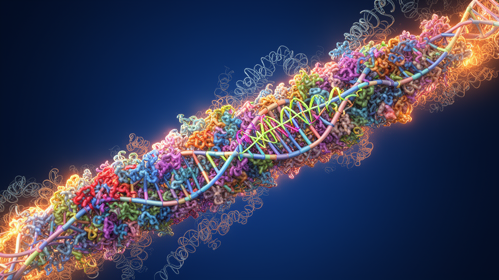
Image Credits: AI Generated
DOI: 10.1007/s11357-026-02588-y
Keywords: mitochondria, inflammaging, cellular senescence, MiDAS, mitochondrial DNA, SASP, cGAS-STING, NLRP3 inflammasome, mitophagy, regulated cell death, reactive oxygen species, ageing




















 English (US) ·
  French (CA) ·