

 1 hour ago
6
1 hour ago
6
PROTECT YOUR DNA WITH QUANTUM TECHNOLOGY
Orgo-Life the new way to the future Advertising by AdpathwayA team of researchers has used living human brain tissue to reveal how deep brain stimulation changes the activity of individual brain cells and activates genetic programs linked to memory and brain plasticity. The study, published in Nature, offers one of the clearest molecular views yet of how electrical stimulation may influence the human brain—and points toward possible future treatments for cognitive decline.
Deep brain stimulation, or DBS, is a clinical technique in which implanted electrodes deliver controlled electrical impulses to specific regions of the brain. It is already used to reduce symptoms of Parkinson’s disease and has also been approved or investigated for conditions including obsessive-compulsive disorder, epilepsy and severe movement disorders. More recently, scientists have begun testing whether carefully designed stimulation could improve memory or slow aspects of cognitive deterioration.
Although the clinical potential of DBS has attracted growing attention, researchers have known relatively little about what happens inside different types of human brain cells when the electrical pulses are delivered. Most previous work has relied on animals, laboratory-grown cells or organoids. Those systems are valuable, but they cannot fully reproduce the organization, genetic diversity and disease history of an adult human brain. The new investigation is believed to be the first to reproduce DBS-like electrical patterns in living human brain tissue outside the body while simultaneously examining the genetic response of individual cells.
The samples came from the temporal cortex, a region on the sides of the brain’s outer layer that plays a central role in memory, language and other cognitive processes. The tissue was donated by neurosurgery patients and maintained under laboratory conditions for several days. While the samples remained viable, the researchers exposed them to electrical stimulation designed to resemble patterns used in deep brain stimulation. This approach allowed the team to examine immediate cellular responses in human tissue that had recently been part of a functioning brain.
One of the most striking observations was a rise in synchrony among brain cells. Neurons communicate through precisely timed electrical and chemical signals, and coordinated activity across groups of neurons is thought to help the brain encode and retrieve memories. By measuring electrical behavior in the tissue, the researchers found that stimulation encouraged cells to communicate in a more synchronized pattern. The result does not prove that the isolated tissue formed new memories, but it reproduces a type of coordinated activity strongly associated with memory formation in living brains.
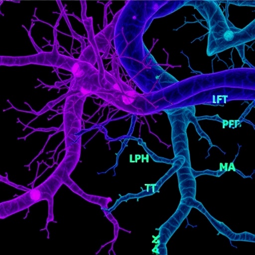
The researchers then examined how stimulation altered gene expression at the level of individual cells. They isolated nuclei from the tissue and used single-cell genetic analysis to determine which genes were active in different cell populations. This technique can distinguish molecular responses that would be hidden in an average measurement of an entire tissue sample. Instead of producing one uniform reaction, the stimulation triggered distinct genetic programs in different types of cells.
Neurons activated groups of genes associated with communication, adaptability and the ability to change their connections. These processes are fundamental to synaptic plasticity, the biological capacity through which neural circuits strengthen, weaken or reorganize in response to experience. Such changes are considered essential for learning and memory. The findings suggest that electrical stimulation may influence memory-related function not only by changing the immediate firing patterns of neurons, but also by altering the molecular machinery that enables cells to adapt over time.
The study also highlighted the response of astrocytes, non-neuronal support cells that help maintain the brain’s chemical environment, provide metabolic assistance and regulate communication between neurons. Astrocytes are increasingly recognized as active participants in information processing rather than passive structural supports. In the stimulated tissue, they switched on their own characteristic genetic programs, indicating that DBS may affect the broader cellular ecosystem of the brain. These responses could help explain why stimulation has effects that extend beyond the neurons directly exposed to an electrical field.
Importantly, the researchers observed comparable molecular patterns in tissue collected from patients who had received brain stimulation before surgery. This comparison suggests that the laboratory findings were not simply an artifact of exposing detached tissue to electricity. Some of the same cellular responses appeared to occur in the human brain during treatment. However, the scientists caution that the experiments represent a limited window into a complex process. The tissue was studied for a relatively short period, and the long-term consequences of repeated stimulation remain unknown.
By identifying which genes are activated in which cell types, the work could help researchers make DBS more precise. Genetic targets might eventually be paired with stimulation to amplify beneficial effects or reduce unwanted ones. For example, drugs could be designed to influence molecular pathways that support plasticity, while electrical parameters could be adjusted to recruit specific neural circuits. Such a combined strategy could be relevant to memory disorders and neurodegenerative diseases, although significant research is still required before these possibilities can be tested clinically.
The researchers emphasize that important questions remain. Future studies will need to determine how long the genetic changes persist, how stimulated cells communicate with neighboring populations and whether similar responses occur in deeper brain structures that are difficult to obtain from living donors. The work nevertheless provides a rare, direct look at human brain tissue responding to therapeutic electrical patterns. By connecting synchronized neural activity with cell-specific genetic changes, it brings scientists closer to understanding—and potentially improving—the way brain stimulation is used to preserve cognition.
Subject of Research: Human tissue samples
Article Title: Stimulation modulates gene-linked cell assemblies in the human brain
News Publication Date: 5-Aug-2026
Web References: https://neurobio.ucla.edu/people/genevieve-konopka-phd
References: Nature
Keywords: Deep brain stimulation, neuroscience, human brain tissue, brain stimulation, memory, cognitive decline, gene expression, astrocytes, neurons, synaptic plasticity, neurology, neurosurgery, Parkinson’s disease, Alzheimer’s disease
Tags: brain plasticity and memory enhancementDBS impact on cognitive declinedeep brain stimulation molecular mechanismseffects of electrical stimulation on brain tissueselectrical stimulation effects on neuronsgenetic programs activated by DBShuman brain cell activityhuman brain tissue gene activitymolecular insights into brain stimulationneural response to electrical impulsesneurological treatment advancementsunderstanding neuronal changes in human brain

















 English (US) ·
  French (CA) ·