

 1 month ago
72
1 month ago
72
PROTECT YOUR DNA WITH QUANTUM TECHNOLOGY
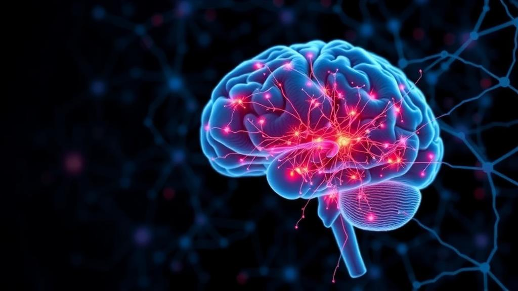
Orgo-Life the new way to the future Advertising by AdpathwayIRVINE, Calif., Aug. 20, 2026 — The human cerebral cortex is often portrayed as a single folded sheet of neural tissue, but its remarkable abilities depend on a much more precise arrangement. During embryonic development, the cortex is divided into areas with distinct molecular identities and developmental trajectories. Regions toward the front of the brain eventually contribute to functions including planning, decision-making, language and social behavior, while areas toward the back become specialized for processing sensory information, including vision. Scientists refer to this process as cortical arealization, and researchers at the University of California, Irvine, have now developed a stem cell-based system that reproduces a key part of it in the laboratory.
In a study published in Cell Stem Cell, the UC Irvine-led team created human neocortical organoids with a defined anteroposterior identity, meaning that the tissues acquired molecular characteristics associated with either the front or the back of the developing cerebral cortex. Brain organoids are three-dimensional tissues produced from human pluripotent stem cells. Under carefully controlled conditions, these cells self-organize into structures that reproduce selected features of early human brain development, including neural progenitor cells, immature neurons and layered patterns of gene activity. However, conventional organoids often develop as a mosaic of randomly specified regions, making it difficult to determine where a particular cell or defect would belong in the developing brain.
The new approach addresses that limitation by applying developmental signals at an early stage of organoid formation. In the embryo, secreted morphogens act as positional cues, forming chemical gradients that tell cells where they are located and which regional programs they should activate. The UC Irvine researchers used selected signaling molecules to bias organoids toward front-like or back-like cortical identities. These signals influenced the expression of transcription factors and other genes that regulate neural cell fate, effectively providing the growing tissue with a reproducible biological coordinate system. Rather than producing an anatomically complete brain, the method generates cortical tissue with a more clearly defined regional identity that can be compared across experiments.
To test whether the engineered tissues truly resembled corresponding regions of the developing human cortex, the researchers examined individual cells using high-throughput molecular profiling. Their analysis included more than 200,000 cells, allowing them to compare gene-expression patterns across organoids and with reference data from prenatal human brain tissue. The results showed that front-directed and back-directed organoids expressed distinct molecular programs associated with their intended locations. These programs were not limited to a single marker; instead, they involved coordinated patterns across populations of neural progenitors and developing neurons. The findings indicate that morphogen-guided organoids can capture important aspects of human cortical regionalization that are largely absent from unpatterned models.
“Brain organoids have become powerful tools for studying human development, but the human brain is a highly organized space,” said lead author Momoko Watanabe, an assistant professor of anatomy and neurobiology at the UC Irvine School of Medicine and a faculty member of the Sue & Bill Gross Stem Cell Research Center. “By introducing regional identity into these models, we can begin asking questions about development and disease that were difficult to address with conventional organoids.” The ability to assign tissue a front-to-back identity could allow scientists to investigate how neighboring cortical areas acquire different cell types, establish distinct connectivity and respond differently to genetic or environmental disruption.
The researchers then applied the platform to fragile X syndrome, a genetic neurodevelopmental condition and the leading inherited cause of intellectual disability. Fragile X syndrome is caused by changes involving the FMR1 gene and is frequently associated with autism spectrum disorder, altered learning and memory, and other neurological features. The team compared regionalized organoids generated from donors with and without the condition. Their focus included SOX4 and SOX11, two transcription factors involved in neural development whose levels normally differ between front and back cortical tissue. In organoids derived from unaffected donors, the expected regional contrast in SOX4 and SOX11 was consistently maintained. In fragile X syndrome organoids, however, that contrast was substantially reduced.
The result suggests that fragile X syndrome may influence more than the behavior of individual neural cells. It may also alter the molecular distinctions that normally organize developing cortical regions. Importantly, the broad front-to-back identity of the engineered organoids remained detectable, indicating that the disorder did not simply erase regionalization altogether. Instead, a more specific developmental relationship between cortical position and gene regulation appeared to be weakened. The researchers note that similar flattening of SOX4 and SOX11 differences has been reported in donated brain tissue from people with autism. The organoid findings do not demonstrate that disrupted cortical patterning causes autism, but they provide a controlled human model in which the relationship can be examined experimentally.
Regionalized organoids could be valuable because many neurological and neurodevelopmental disorders do not affect every part of the brain equally. A model that preserves positional information may help researchers ask not only which genes, pathways or cell types are altered, but also where and when those alterations first emerge. Scientists could use the system to compare disease-associated variants, study the effects of environmental exposures during early development, or test whether candidate treatments restore regional molecular differences. Because the organoids are derived from human stem cells, they may also capture aspects of cortical development that differ between humans and commonly used animal models, while still allowing experiments that would be impossible to perform directly in a developing human brain.
The study also demonstrates how developmental biology and tissue engineering can be combined to make organoids more reproducible and informative. Morphogen exposure does not recreate the full architecture of the fetal cortex, and the resulting tissues remain simplified models rather than miniature brains. They lack many features of a complete developing nervous system, including the full range of long-distance connections, vascular interactions and influences from other cell types and organs. Even so, assigning a defined anteroposterior identity represents a significant step toward building more organized human neural tissues. The UC Irvine team, whose collaborators include researchers in stem cell biology, developmental biology, mathematics and computational science, plans to use the platform to explore how regional identities interact with cell maturation and disease mechanisms.
The work, titled “Morphogen-guided neocortical organoids with anteroposterior areal identity,” was conducted by investigators from UC Irvine’s School of Medicine, School of Physical Sciences, Charlie Dunlop School of Biological Sciences, Sue & Bill Gross Stem Cell Research Center and NSF-Simons Center for Multiscale Cell Fate Research, with collaboration from the University of Pennsylvania. Support came in part from the National Institutes of Health, National Science Foundation, California Institute for Regenerative Medicine, Simons Foundation and FRAXA Research Foundation. By giving lab-grown human cortical tissue a defined developmental sense of place, the researchers have created a model that could make some of the brain’s earliest organizational decisions visible—and potentially reveal how those decisions go awry in disease.
Subject of Research: Human cortical development, cortical arealization, neocortical organoids and fragile X syndrome
Article Title: Morphogen-guided neocortical organoids with anteroposterior areal identity
News Publication Date: Aug. 20, 2026
Web References: Cell Stem Cell article; University of California, Irvine News
References: Watanabe, Momoko, et al. “Morphogen-guided neocortical organoids with anteroposterior areal identity.” Cell Stem Cell.
Keywords: brain organoids, cerebral cortex, cortical arealization, human stem cells, fragile X syndrome, autism research, morphogens, neurodevelopment, cortical development, regenerative medicine
Tags: 3D human brain tissue modelsadvances in neuroscience researchbrain organoidscortical arealizationembryonic brain developmentfunctional brain region differentiationhuman cerebral cortex developmentin vitro brain tissue engineeringmolecular identity of brain regionsneural tissue self-organizationspatial awareness in lab-grown brainsstem cell-derived neocortical models


















 English (US) ·
  French (CA) ·