

 1 hour ago
8
PROTECT YOUR DNA WITH QUANTUM TECHNOLOGY
Orgo-Life the new way to the future Advertising by AdpathwayA surgical team has successfully managed a rare and complex case of rectal perforation caused by a migrated intrauterine device that remained hidden while a second contraceptive implant sat in its normal intrauterine position, according to a clinical case report published in the journal BMC Women’s Health. By detailing the patient’s presentation and surgical treatment, the report highlights the critical diagnostic vulnerabilities associated with misplaced intrauterine contraceptives.
Translocation of an intrauterine contraceptive device beyond the uterine cavity and into the gastrointestinal tract, specifically the rectum, represents an uncommon but potentially life-threatening complication in gynecological practice. According to the study authors, the simultaneous presence of two distinct intrauterine devices within a single patient—one retaining its appropriate placement inside the uterine cavity while another silently migrates into adjacent pelvic structures—is an even more exceptional occurrence that creates an intricate diagnostic challenge.
The case involved a 47-year-old woman, identified as gravida 4, para 2, who presented to healthcare providers with a persistent one-month history of loose stools. A comprehensive review of her gynecological history documented multiple contraceptive placements spanning more than two decades. Her very first intrauterine device, a Mutile model, had been inserted more than 20 years earlier but was subsequently presumed to have been spontaneously expelled after it could no longer be located. Clinicians later placed a second device, which was subsequently removed following episodes of abnormal vaginal bleeding. Approximately ten years prior to her clinical presentation, a third device, designated as an AiMu model, was inserted and verified as correctly positioned inside the uterine cavity.
A series of diagnostic imaging assessments and endoscopic procedures ultimately uncovered the presence of the long-lost foreign object and demonstrated the true extent of the structural injury. During a colonoscopy examination, physicians discovered a black metallic foreign body embedded directly within the rectal wall at a distance of 10 centimeters from the anal verge, accompanied by localized abscess formation in the surrounding tissue. Further cross-sectional imaging with computed tomography confirmed the location of the dense metallic foreign object, visualizing it within the mid-lower rectum at a distance of 66 millimeters from the anal verge. In addition, a pelvic ultrasound examination clearly demonstrated the dual-IUD configuration, revealing that two separate contraceptive devices remained inside the patient’s pelvis simultaneously.
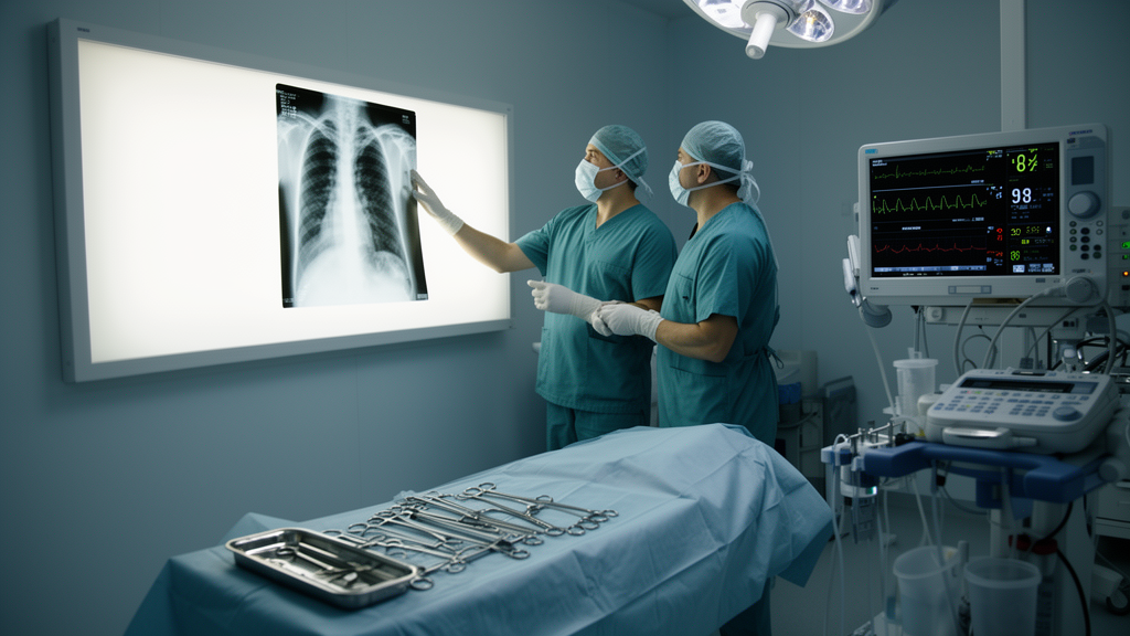
To address the dual foreign bodies and manage the associated colorectal injury, the medical team devised a combined surgical intervention utilizing both hysteroscopic and laparoscopic techniques. During the first phase of the operation, surgeons accessed the uterine cavity hysteroscopically to safely extract the normally positioned AiMu device. Following the successful laparoscopic retrieval of the foreign object, the surgical team completed a primary surgical repair of an anterior rectal perforation that measured 1.0 by 0.3 centimeters.
Following the combined surgical intervention, the patient experienced a smooth and uneventful recovery under close hospital observation. Throughout the subsequent outpatient follow-up period, the protective terminal ileostomy remained healthy, viable, and fully functional, ensuring proper intestinal diversion.
Reflecting on the findings of this case, the authors emphasized important clinical lessons regarding foreign body localization and contraceptive surveillance. The authors concluded that the simple failure to visualize an intrauterine device during routine pelvic ultrasound must not be taken as definitive confirmation of spontaneous expulsion. When the precise anatomical localization of an intrauterine device remains uncertain, or whenever extrauterine migration is suspected, the researchers advocate that computed tomography represents an appropriate and necessary diagnostic modality to clarify the clinical picture.
Subject of Research: Laparoscopic management of rectal perforation caused by a migrated intrauterine device coexisting with an intrauterine implant
Article Title: Laparoscopic management of rectal perforation caused by a migrated intrauterine device with a coexisting intrauterine device: a case report
Article References: Huang, R., Deng, J., Xu, L., Wan, L., & Chen, H. (2026). Laparoscopic management of rectal perforation caused by a migrated intrauterine device with a coexisting intrauterine device: a case report. BMC Women's Health. https://doi.org/10.1186/s12905-026-05018-5
Image Credits: AI Generated
DOI: 10.1186/s12905-026-05018-5
Keywords: intrauterine device migration, rectal perforation, laparoscopic retrieval, hysteroscopic removal, protective ileostomy, terminal ileostomy, ileostomy reversal, ectopic IUD, Mutile IUD, AiMu IUD, pelvic ultrasound, computed tomography, colonoscopy, rectal foreign body, visceral migration, dual IUD, BMC Women's Health



















 English (US) ·
 French (CA) ·