

PROTECT YOUR DNA WITH QUANTUM TECHNOLOGY
Orgo-Life the new way to the future Advertising by Adpathway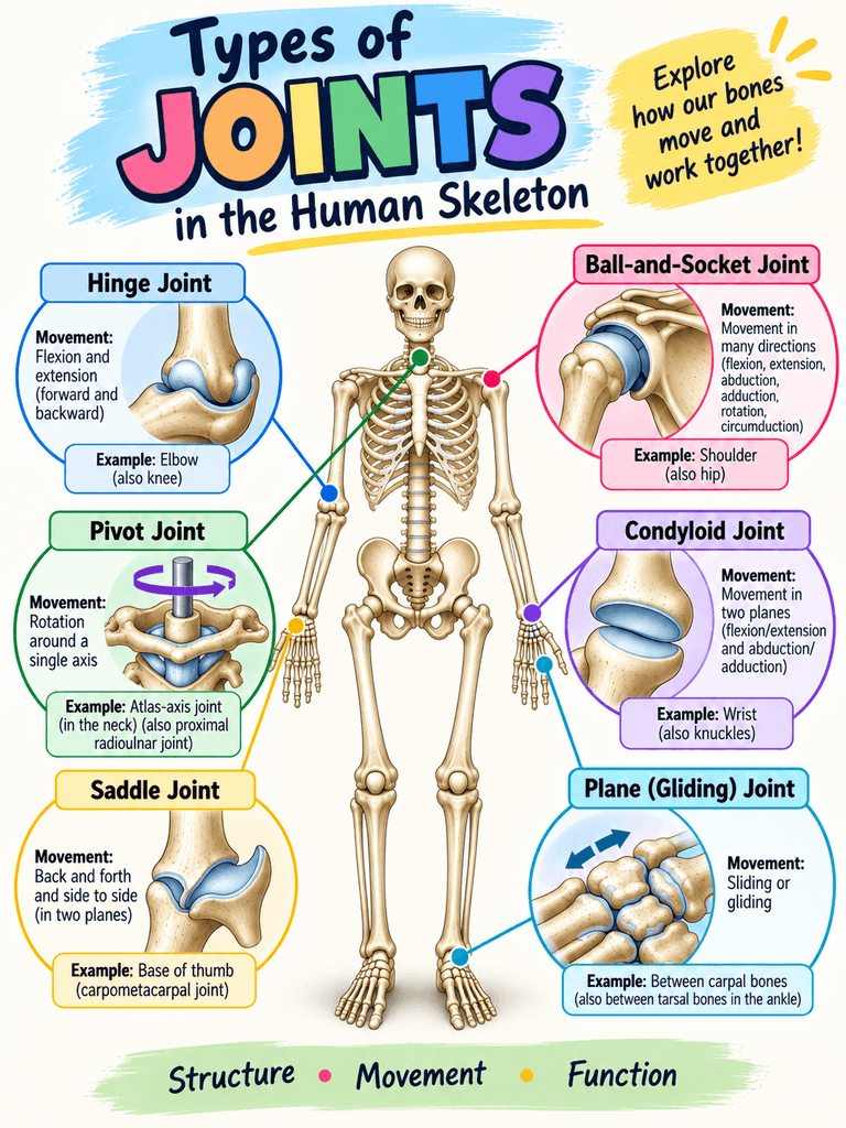
When students first learn the skeletal system, usually they focus their attention on naming the bones. Knowing the names of the bones is important, but bones are only part of the story. Joints are what allow the skeleton to move. You can also discuss sports injuries that are common to joints, like an ACL tear or a dislocated shoulder. Students love to share stories about their injuries, often in gruesome detail!
The Types of Joints in the Human Skeleton worksheet helps anatomy students move beyond bone identification by examining where bones meet, how joints are classified, and how the structure of a joint determines its movement.
Students can label the joints by referencing their classroom slides, which has a section on the joints. These slides cover the entire unit, which includes the microscopic structure of bones, types of bones, bone growth, and disorders of the skeletal system.
Exploring the Six Types of Synovial Joints
The main focus of the activity is the six types of synovial joints. Students identify examples on the human skeleton and compare the movements each type allows.
Pivot joints permit rotation around an axis. Students can observe this movement when they turn their heads from side to side at the atlas-axis joint or examine rotation at the proximal radioulnar joint.
Hinge joints primarily allow flexion and extension, much like the movement of a door hinge. The elbow is a classic example, while the knee is often described as a modified hinge joint.
Ball-and-socket joints provide movement in multiple directions as well as rotation. The shoulder and hip are the major examples. Their large range of motion makes them especially useful for comparing joint structure and function.
Saddle joints allow movement in two planes. The joint at the base of the thumb is an important example and contributes to the remarkable mobility of the human thumb.
Condyloid joints (also called ellipsoid) also permit movement in two planes, including flexion, extension, abduction, and adduction. The wrist provides an accessible example for students to explore.
Plane joints (also called gliding), allow relatively flat bone surfaces to slide past one another. These joints occur between many of the carpal bones of the wrist and tarsal bones of the ankle.
In the Classroom
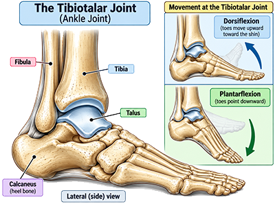
This activity works particularly well when students have access to an articulated classroom skeleton. They can also use individual models showing joints like the knee or the ankle. Instead of identifying joints only from textbook diagrams, students can locate structures directly on a model.
The worksheet includes a skeleton diagram where students identify the locations of different synovial joints. Close-up illustrations help students visualize the shapes of the articulating surfaces and connect those shapes to the movements they permit.
You can also turn joint identification into a movement activity. Ask students to flex their elbow, rotate their head, circle their arm at the shoulder, move their thumb across their palm, or flex their ankle. Students can then determine which joint is moving and classify it.
The Types of Joints in the Human Skeleton activity includes identification of the six synovial joint types. Students label the types of joints on an image. Then they match the names of joints with the type. For example, the knee is a hinge joint. Finally, students consider different movements of the body and what joints are involved. When you bend your elbow, what kind of joint is responsible? (Answer: it’s a hinge joint, just like the knee!)
The resource can be used as an introduction to joints, as part of a skeletal-system unit, or as a follow-up activity after students have learned the major bones of the human skeleton. It also works well as a station activity where students rotate between anatomical models and identify joints as they complete the worksheet.
Related Resources
The Skeletal System Unit Guide – includes all of my resources for teaching the skeletal unit
Coloring the Human Skeleton – students use their notes or other resources to identify bones and color a skeleton
Anatomy of the Knee – focus on the ligaments of the knee joint, like the anterior cruciate ligament (ACL)
Case Study: Ehlers-Danlos Syndrome – explores how a genetic disorder affects the joints



















 English (US) ·
  French (CA) ·