

 1 hour ago
3
PROTECT YOUR DNA WITH QUANTUM TECHNOLOGY
Orgo-Life the new way to the future Advertising by AdpathwayA longitudinal pediatric case series has revealed that endocrine deficiencies and structural pituitary abnormalities in ulnar-mammary syndrome can exhibit divergent evolutionary trajectories, including spontaneous resolution over time. Ulnar-mammary syndrome, designated MIM 181450, represents a rare autosomal-dominant developmental condition characterized by incomplete penetrance that impacts multiple organ systems. Clinically, the disorder manifests with upper limb malformations, mammary and apocrine gland hypoplasia, dental crowding or malocclusion, abnormal hair distribution, and genital anomalies. Additional reported features include cardiac defects, palatal anomalies, and endocrine impairments, with affected males frequently displaying delayed pubertal development and genital hypoplasia. Molecularly, ulnar-mammary syndrome arises from haploinsufficiency of the TBX3 gene, a dosage-sensitive transcriptional regulator essential for embryonic development. While early clinical evaluations often identify growth failure and pubertal delay, systematic long-term observational data tracking these hormone axes through late adolescence have remained scarce.
This knowledge gap hinders clinicians from determining whether presenting endocrine deficits in ulnar-mammary syndrome represent permanent endocrine lesions or transient maturational delays. Distinguishing between reversible conditions, such as constitutional delay of growth and puberty, and permanent endocrinopathies, such as isolated hypogonadotropic hypogonadism, requires prolonged clinical follow-up. To clarify this natural history, pediatric endocrinologists at the Children’s Hospital of Soochow University in Suzhou, China, conducted an extended longitudinal evaluation of two adolescent male patients diagnosed at twelve years of age. By integrating auxological monitoring, serial provocative hormonal testing, and repeated pituitary magnetic resonance imaging with in vitro minigene assays and structural modeling, the researchers documented the longitudinal trajectory of pituitary function and pubertal development. Their findings demonstrate that initial pituitary hypoplasia and growth hormone deficiency can resolve spontaneously in some individuals, whereas other patients exhibit progressive hypothalamic-pituitary structural abnormalities.
The first patient, designated Patient 1, presented initially at twelve years and eight months of age with short stature, delayed pubertal onset, and typical dysmorphic features. His baseline clinical presentation, originally described in a Chinese-language report, included distinct facial dysmorphism, mammary and apocrine gland hypoplasia, prepubertal genitalia, and a height of 148.9 centimeters. Diagnostic evaluation documented growth hormone deficiency on provocative testing accompanied by reduced serum insulin-like growth factor 1 concentrations. A gonadotropin-releasing hormone stimulation test showed a prepubertal response, while neuroimaging demonstrated anterior pituitary hypoplasia on magnetic resonance imaging. These clinical and biochemical findings prompted an initial diagnostic suspicion of isolated hypogonadotropic hypogonadism with concurrent growth hormone deficiency. The current investigation reports previously unpublished longitudinal follow-up data from age thirteen to sixteen years, tracing his endocrine trajectory through pubertal completion following recombinant human growth hormone treatment.
Whole-exome sequencing performed in Patient 1 identified a heterozygous de novo frameshift variant within the TBX3 gene, specifically c.711del (p.N238Mfs*4). Sanger sequencing confirmed the presence of this deletion in the patient and its complete absence in both biological parents. Clinical geneticists classified the variant as pathogenic based on established guidelines from the American College of Medical Genetics and Genomics and the Association for Molecular Pathology. The classification met criteria PVS1, designating a frameshift alteration predicted to cause loss of function in a gene where haploinsufficiency represents a definitive disease mechanism; PS2, confirming a de novo event in a proband exhibiting an aligned clinical phenotype and negative family history; and PM2, reflecting complete absence from general population sequencing databases.
Based on confirmed growth hormone deficiency, Patient 1 initiated recombinant human growth hormone therapy at a standard therapeutic dosage, maintaining treatment compliance for approximately two and a half years. Auxological follow-up documented robust linear growth, with stature progressing from 148.9 centimeters at baseline to 150.0 centimeters at age thirteen, 162.0 centimeters at age fourteen, 165.0 centimeters at age fourteen years and six months, and 174.0 centimeters at age fifteen years and eight months. Serial bone age assessments demonstrated steady skeletal maturation, advancing from thirteen years at chronological age thirteen to thirteen years and six months at age fourteen years and six months, and reaching fourteen years at chronological age sixteen. At age sixteen, his predicted adult height reached 180.0 centimeters, significantly surpassing his mid-parental target height of 165.5 centimeters and confirming favorable long-term auxological catch-up during clinical follow-up.
Longitudinal monitoring revealed spontaneous pubertal onset in Patient 1 at fifteen years of age, evidenced by bilateral testicular enlargement. By age fifteen years and eight months, shortly after growth hormone therapy concluded, external genitalia progressed to Tanner stage G3 with testicular volumes of 15 milliliters and pubic hair at Tanner stage P2. Repeated gonadotropin-releasing hormone provocation testing showed robust central hypothalamic-pituitary-gonadal axis activation, with peak luteinizing hormone rising to 15.64 international units per liter, peak follicle-stimulating hormone reaching 7.41 international units per liter, and baseline testosterone measuring 4.82 nanomoles per liter. Furthermore, provocative testing using arginine and clonidine at age sixteen demonstrated a normalized peak growth hormone level of 8.2 micrograms per liter, while cranial magnetic resonance imaging revealed a normal anterior pituitary gland measuring 4.69 millimeters in height. These outcomes demonstrated the resolution of pituitary hypoplasia and growth hormone deficiency, prompting diagnostic revision from isolated hypogonadotropic hypogonadism to constitutional delay of growth and puberty.
In contrast to Patient 1, Patient 2 exhibited persistent, progressive hypothalamic-pituitary dysfunction. Born at term via cesarean delivery with a birth weight of 3,400 grams and length of 50 centimeters to healthy non-consanguineous parents, his medical history included surgical repair of an inguinal hernia at age three and orchiopexy for cryptorchidism at age five. At ten years and ten months of age, with a height of 134.6 centimeters, he was diagnosed with growth hormone deficiency at another facility based on a peak stimulated growth hormone of 5.13 micrograms per liter and pituitary height of 3.0 millimeters on magnetic resonance imaging. Outside growth hormone therapy increased his height to 145.5 centimeters by October 2023, when treatment was discontinued by his family. Presenting to the study hospital at twelve years and six months, his height was 147.0 centimeters with prepubertal genitalia, mammary and apocrine gland hypoplasia, dental malocclusion, and absent axillary hair, without facial dysmorphism.
Diagnostic re-evaluation in Patient 2 demonstrated an intricate endocrine trajectory. Following a washout period exceeding five months, a repeat provocative growth hormone test at twelve years and six months yielded a normal peak level of 14.1 micrograms per liter; however, documented growth arrest prompted the re-initiation of growth hormone therapy at 0.13 international units per kilogram daily. At thirteen years and eight months, genitalia remained prepubertal with low gonadotropins and basal testosterone below 0.24 nanomoles per liter. Pituitary magnetic resonance imaging revealed progressive hypoplasia measuring 2.5 millimeters and non-visualization of the neurohypophysis, consistent with pituitary stalk interruption. Because testicular volume remained 2 milliliters at fourteen years and two months, clinicians added subcutaneous human chorionic gonadotropin and human menopausal gonadotropin. After five months of gonadotropin therapy, bilateral testicular volume increased to 4 milliliters and testosterone rose to 8.34 nanomoles per liter, confirming intact Leydig cell function. Following dose titration of growth hormone to 0.152 international units per kilogram daily, height reached 161.7 centimeters at fourteen years and eight months.
Genetic analysis in Patient 2 revealed a de novo heterozygous splice-site variant in TBX3, identified as c.658-3C>G. Absent from population databases, the variant was classified as likely pathogenic based on American College of Medical Genetics criteria PS3_Strong, PS2, PM2, and PP3. To experimentally evaluate the splicing consequences, researchers developed an in vitro minigene assay. Genomic fragments spanning exon 2, intron 2, exon 3, and partial intron 3 were cloned into pcMINI-N vectors and transfected into HeLa and HEK293T cells. Reverse transcription polymerase chain reaction analysis demonstrated that the wild-type construct generated the expected 575-base-pair transcript, whereas the mutant construct produced two aberrant transcripts. Band b exhibited retention of eighty base pairs of intronic sequence upstream of exon 3, predicting a truncated 229-amino-acid protein (p.Thr220Tyrfs*11) or nonsense-mediated decay. Band c displayed complete skipping of exon 3 (c.658_804del), predicting an in-frame deletion of forty-nine amino acids (p.Thr220_Lys268del) within the T-box domain.
Protein structural modeling utilizing SWISS-MODEL and PyMOL visualization illustrated the conformational consequences of the predicted mutant proteins. Truncation and internal deletion were predicted to alter the tertiary architecture of the conserved T-box domain and reduce overall protein stability, aligning with a loss-of-function mechanism. During embryonic development, TBX3 expression occurs within Rathke’s pouch and the infundibulum, establishing its essential role in hypothalamic-pituitary organogenesis. Furthermore, TBX3 regulates alternative splicing via its C-terminal domain, whereas mutations in the 5-prime portion of the T-box domain predominantly impair this splicing function. However, the study authors explicitly noted that downstream effects on translation efficiency, intracellular stability, DNA binding, and transcriptional regulation were not experimentally quantified in living cells, leaving these functional implications predicted rather than directly measured. This distinction between direct splicing observations and theoretical protein consequences remains an important boundary in interpreting the molecular findings.
The divergent outcomes in these two patients emphasize that endocrine deficits in ulnar-mammary syndrome vary across individuals and over time, cautioning clinicians against drawing premature prognostic conclusions. Patient 1 demonstrated complete normalization of growth hormone secretion, pubertal progression, and resolution of pituitary hypoplasia, showing that early presentation mimicking isolated hypogonadotropic hypogonadism can represent constitutional delay of growth and puberty. Conversely, Patient 2 exhibited progressive hypothalamic-pituitary pathology, with pituitary stalk interruption requiring gonadotropin replacement. These findings demonstrate that baseline neuroimaging does not always correlate with endocrine function, highlighting the necessity of long-term hormonal surveillance of growth hormone and gonadotropic axes until final adult height and sexual maturity. The principal limitations of the investigation comprise its retrospective design and small sample size of two patients, underscoring the need for larger prospective cohorts to validate these clinical patterns.
Subject of Research: Endocrine trajectories and TBX3 variants in pediatric ulnar-mammary syndrome
Article Title: Case Report: Long-term follow-up in ulnar-mammary syndrome caused by novel TBX3 variants: a case series with longitudinal endocrine follow-up
Article References: Wang, X., Chen, X., Wu, H., Xie, R., Chen, T., Chen, L., & Wang, F. (2026). Case Report: Long-term follow-up in ulnar-mammary syndrome caused by novel TBX3 variants: a case series with longitudinal endocrine follow-up. Frontiers in Pediatrics, 14, Article 1931471. https://doi.org/10.3389/fped.2026.1931471
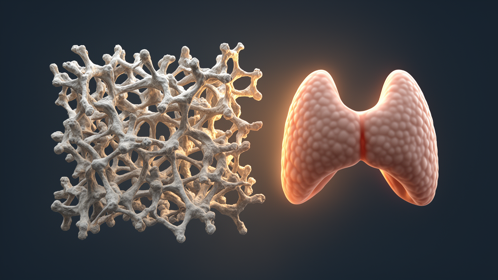
Image Credits: AI Generated
DOI: 10.3389/fped.2026.1931471
Keywords: ulnar-mammary syndrome, TBX3, growth hormone deficiency, pituitary hypoplasia, delayed puberty, constitutional delay of growth and puberty, hypogonadotropic hypogonadism, minigene assay, splice-site mutation, frameshift variant, pituitary stalk interruption, recombinant human growth hormone, gonadotropin-releasing hormone test, T-box domain, pediatric endocrinology, auxological assessment, Rathke pouch



















 English (US) ·
 French (CA) ·