

 4 hours ago
5
PROTECT YOUR DNA WITH QUANTUM TECHNOLOGY
Orgo-Life the new way to the future Advertising by AdpathwayPrimary microglia deficient in either the cell surface receptor TREM2 or its transmembrane signaling adaptor TYROBP exhibit fundamentally distinct downstream intracellular signaling trajectories, cellular morphologies, and gene expression cascades despite functioning within the same canonical receptor complex. While triggering receptor expressed on myeloid cells 2 and TYRO protein tyrosine kinase binding protein physically couple in microglial membranes to coordinate immune responses, their individual genetic ablation produces divergent functional phenotypes rather than identical cellular deficits. By comparing isolated primary microglia from wild-type, Trem2 knockout, and Tyrobp knockout neonatal mice, researchers demonstrated that the loss of either component causes baseline signaling dysregulation, characterized by elevated basal levels of phosphorylated ERK and AKT. However, subsequent exposure to inflammatory or Alzheimer’s disease-associated pathological stimuli revealed that microglia lacking TYROBP mount markedly different biochemical and transcriptomic responses than cells lacking TREM2, showing that these two partners execute non-redundant regulatory roles in microglial homeostasis and activation.
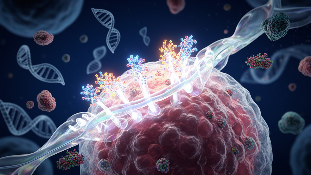
In the mammalian brain, microglia serve as primary resident immune cells capable of shifting across diverse activation states that govern neuroprotection, neurotoxicity, cytokine production, cell survival, proliferation, and phagocytosis. Under homeostatic conditions, microglial cell bodies extend ramified processes to survey the local neural parenchyma. In neuropathological states such as Alzheimer’s disease, microglial surface receptors, including TREM2 and CD33, become activated in response to pathological ligands, including amyloid beta peptides, cellular debris, lipopolysaccharides, and various lipoproteins. Upon ligand engagement, the short intracellular domain of TREM2, which lacks intrinsic enzymatic activity, relies on its physical association with the transmembrane adaptor TYROBP. This interaction drives phosphorylation of the immuno-tyrosine activation motif within TYROBP by Src family kinases. Phosphorylated ITAM domains subsequently generate docking platforms for spleen tyrosine kinase, triggering downstream signaling cascades that recruit mitogen-activated protein kinase, phosphatidylinositol-3 kinase, phospholipase C gamma, and VAV protein families to guide microglial survival and immune activity.
To directly contrast the signaling networks regulated downstream of TREM2 and TYROBP within an isolated microglial setting, the research team harvested primary microglia from newborn C57BL/6 wild-type, Trem2 knockout, and Tyrobp knockout mouse pups between postnatal days 0 and 3. Microglial cells were isolated from homogenized cortical and hippocampal tissues using CD11b-positive magnetic bead separation via magnetic-activated cell sorting. Cultured primary cells were maintained in supplemented growth media before undergoing defined experimental treatments. The investigative design evaluated microglial responses under resting conditions and following exposure to disease-relevant stimuli, specifically lipopolysaccharide to simulate acute neuroinflammatory signaling, isolated OC-type amyloid beta 42 fibrils, or a mixed preparation containing both A11-type prefibrillar oligomers and OC-type fibrils. The cellular preparations enabled direct interrogation of microglial-intrinsic pathways without confounding secondary signals originating from surrounding astrocytes, oligodendrocytes, or neuronal populations.
Microscopic evaluation of baseline cellular architecture uncovered significant morphological transformations caused by the absence of either signaling partner. Wild-type primary microglia displayed an elongated, ramified morphology with small soma volumes and fine processes typical of resting immune surveillance. In contrast, both Trem2 knockout and Tyrobp knockout cells exhibited noticeably larger cell bodies and shortened, retracted processes at baseline, indicative of an activated basal state. Following lipopolysaccharide challenge, Tyrobp knockout microglia displayed a marked reduction in cell area as measured by CD68 immunostaining and circularity analysis, whereas Trem2 knockout cells exhibited no significant morphological change relative to their baseline. Furthermore, baseline functional assessments revealed that both knockout lines had significantly diminished phagocytic uptake of green fluorescent latex beads compared to wild-type controls. However, upon exposure to lipopolysaccharide or amyloid beta fibrils, Trem2 knockout microglia showed increased bead phagocytosis relative to their baseline, whereas Tyrobp knockout microglia failed to mount an equivalent phagocytic response, highlighting a more rigid functional defect in the absence of TYROBP.
Analysis of cell motility through scratch-wound healing assays further accentuated functional disparities between the genotypes. At baseline, microglial migration did not differ significantly between wild-type and either knockout strain, although Trem2 knockout cells displayed a downward trend compared to Tyrobp knockout cells. When exposed to external challenges, wild-type and Trem2 knockout microglial motility remained largely stable across treatments. Conversely, Tyrobp knockout microglia exhibited a significant decrease in migratory capacity specifically following treatment with isolated OC-type amyloid beta fibrils. These functional assay patterns established that although Trem2 and Tyrobp deficiencies produce comparable baseline impairments in morphological ramification and resting bead phagocytosis, the cells diverge substantially once challenged with immune stimuli. Microglia lacking TYROBP demonstrated greater morphological remodeling following endotoxin exposure but suffered deeper deficits in stimulated phagocytosis and fibril-induced cell motility compared to cells lacking TREM2.
Biochemical characterization through immunoblotting revealed substantial disruptions in baseline and stimulated protein phosphorylation across several key intracellular signaling nodes. Under baseline resting conditions, both Trem2 knockout and Tyrobp knockout microglia exhibited elevated phosphorylation of ERK1/2 and AKT at Thr308 and Ser473 compared to wild-type controls, reflecting anomalous resting kinase activation. Upon stimulation with lipopolysaccharide over time intervals of 5, 15, and 30 minutes, wild-type microglia mounted a time-dependent elevation in phosphorylated ERK. In Trem2 knockout microglia, phosphorylated ERK remained persistently elevated without further stimulation-dependent increases. Strikingly, Tyrobp knockout microglia responded to lipopolysaccharide with a significant, rapid decrease in phosphorylated ERK over time, running completely counter to the wild-type response. Baseline phosphorylation of spleen tyrosine kinase at Tyr525/526 and Tyr352 remained unaltered across all three genotypes, indicating that the aberrant baseline phosphorylation of ERK and AKT was not driven by spontaneous changes in resting Syk phosphorylation levels.
Exposure to Alzheimer’s-relevant amyloid beta proteoforms triggered equally distinctive biochemical trajectories. Treatment with OC-type amyloid beta fibrils for 24 hours produced significant increases in phosphorylated ERK and phosphorylated mTOR in wild-type cells, whereas a mixture of prefibrillar A11 oligomers and OC fibrils failed to elicit comparable increases in wild-type cells. In Trem2 knockout microglia, phosphorylated ERK levels remained elevated and unresponsive to either fibril preparations or oligomer-fibril mixtures. In contrast, Tyrobp knockout microglia exposed to OC-type amyloid beta fibrils displayed a significant reduction in phosphorylated ERK relative to untreated knockout baselines, mirroring the downward shift observed following endotoxin exposure. Phosphorylated mTOR, which trended upward in wild-type microglia after fibril stimulation, remained unchanged in both knockout models. These biochemical findings indicate that baseline disruptions in ERK activation persist even across extended 24-hour incubation periods in serum-free conditions, with Tyrobp-deficient cells consistently deactivating the ERK pathway under conditions that activate it in wild-type cells.
Pharmacological inhibitor assays dissected the dependence of these signaling events on upstream kinase and receptor pathways. Pre-incubating cells with the selective spleen tyrosine kinase inhibitor BAY61-3606 reduced lipopolysaccharide-induced ERK phosphorylation in wild-type cells and lowered ERK phosphorylation across stimulated Trem2 knockout cells. However, in Tyrobp knockout microglia, Syk inhibition caused a significant reduction in phosphorylated ERK under basal conditions, demonstrating that Tyrobp-deficient microglia rely directly on basal Syk activity to maintain their elevated baseline ERK activation. Blocking Toll-like receptor 4 using the small-molecule inhibitor TAK-242 reduced baseline ERK phosphorylation in both knockout models and attenuated lipopolysaccharide-stimulated ERK phosphorylation across all groups, while failing to block fibril-mediated signaling. In contrast, inhibiting protein phosphatases 1 and 2A with okadaic acid augmented lipopolysaccharide-induced ERK phosphorylation exclusively in wild-type microglia, while exerting no significant impact in either knockout group, suggesting that PP1 and PP2A do not mediate the baseline phospho-ERK dysregulation observed in the absence of TREM2 or TYROBP.
High-throughput transcriptomic profiling via bulk RNA sequencing unveiled broad, divergent alterations in baseline microglial gene expression. Compared to wild-type controls at baseline, Trem2 knockout microglia exhibited 1,190 downregulated and 1,253 upregulated differentially expressed genes, whereas Tyrobp knockout microglia exhibited 2,676 downregulated and 2,288 upregulated genes. A direct comparison between Tyrobp knockout and Trem2 knockout microglia at baseline identified 99 downregulated and 77 upregulated transcripts. Trem2 knockout microglia showed significant downregulation of core activation genes such as Spp1, Itgax, Rgs1, and Dok2, alongside upregulation of inflammatory signaling genes including Gbp5, Ifi44l, and Ifi202b. In Tyrobp knockout cells, top downregulated genes were prominently linked to cell migration and phagocytosis, including Mmp12, Atp6v0d2, and Ptger4, while upregulated genes included mediators of cytokine signaling and immune cell interactions such as Htr2c, Cd209g, and Mpzl2.
Targeted examination of microglial transitional signatures demonstrated that both genetic deletions disrupted both homeostatic and disease-associated microglial programs. Transcripts characteristic of homeostatic microglia, including Hexb, Cx3cr1, Ctsd, C1qa, and C1qb, were significantly reduced in both Trem2 knockout and Tyrobp knockout microglia at baseline relative to wild-type controls. Furthermore, Stage 1 disease-associated microglial markers Apoe and B2m were downregulated in both knockout strains, whereas Cstb, Fth1, and Lyz2 were downregulated selectively in Tyrobp knockout microglia. Stage 2 disease-associated markers, including Ctsl, Lpl, Cd9, Ccl6, Itgax, Clec7a, and Lilrb4, were uniformly suppressed in both knockouts, while Cst7 was downregulated and Csf1 was upregulated uniquely in Tyrobp-deficient cells. Canonical pathway analyses highlighted striking oppositions: canonical pathways for Phagosome Formation, FAK signaling, and ERK/MAPK signaling were predicted by positive z-scores to be activated in Trem2 knockout cells but strongly inhibited in Tyrobp knockout cells at baseline.
Transcriptomic responses following 24-hour challenge with lipopolysaccharide, isolated fibrils, or oligomer-fibril mixtures underscored further mechanistic divergence. While wild-type and Trem2 knockout microglia exhibited minimal transcriptional alterations following treatment with the A11/OC amyloid mixture, Tyrobp knockout microglia mounted 1,779 differentially expressed genes, indicating retained responsiveness to mixed proteoforms. Ingenuity Pathway Analysis revealed that following lipopolysaccharide exposure, integrin signaling, actin cytoskeleton signaling, and STAT3 pathways were predicted to be inhibited in Trem2 knockout cells yet activated in Tyrobp knockout cells. Following amyloid fibril treatment, the complement system pathway was inhibited in wild-type and Trem2 knockout cells but activated in Tyrobp knockout cells. These pathway patterns indicate that Trem2 deficiency promotes suppression of immune and cholesterol pathways, whereas Tyrobp deficiency unleashes overactive complement and distinct lipid-signaling programs.
Profiling conditioned cell culture media using high-throughput proximity extension assay proteomics via the Olink mouse exploratory panel confirmed significant differences in secreted proteins. Out of 92 evaluated biomarkers, multiple proteins demonstrated significant genotype-by-treatment interactions. Under basal conditions, Trem2 knockout microglia secreted higher concentrations of EpCAM, a regulator of cell adhesion and survival, whereas Tyrobp knockout microglia released higher levels of the transcription factor FLI1 compared to wild-type controls. Exposure to the amyloid mixture markedly elevated extracellular levels of the intracellular protein TNNI3 exclusively in Trem2 knockout media, suggesting altered cell membrane permeability or vesicle release. Extracellular matrix-associated proteins, including WISP1 and MATN2, were specifically decreased in Tyrobp knockout media following amyloid mixture treatment, and S100a4 decreased after lipopolysaccharide exposure. Additionally, anti-inflammatory interleukin-10 secretion in response to lipopolysaccharide was significantly blunted in Trem2 knockout media compared to wild-type and Tyrobp knockout cultures, aligning with an intrinsically heightened pro-inflammatory secretome.
The observed divergence between TREM2 and TYROBP deficiencies illustrates that although these two proteins form an obligate surface receptor complex, their downstream regulatory functions diverge across homeostatic maintenance and active neuroinflammatory states. Methodological considerations must contextualize these findings, as the study utilized primary neonatal microglia cultured in vitro, where baseline expression of adult homeostatic markers such as Tmem119, P2ry12, and Sall1 is known to decline relative to adult in situ microglia. Nonetheless, because wild-type, Trem2 knockout, and Tyrobp knockout primary cells were isolated and cultured under identical standardized conditions, the observed signaling and proteomic discrepancies reflect authentic genotype-dependent differences rather than culture artifacts. Furthermore, because TYROBP serves as a shared signaling adaptor for alternative microglial receptors including SIRPβ1 and TREM1, its deletion disrupts broader immune networks beyond the canonical TREM2 axis. Ultimately, these experimental findings reveal that TREM2 and TYROBP are functionally non-redundant, providing foundational insights into how distinct receptor-adaptor lesions alter microglial physiology in Alzheimer’s disease and Nasu-Hakola disease.
Subject of Research: Differential downstream intracellular signaling, morphology, and gene expression in microglia lacking TREM2 or TYROBP
Article Title: Differential downstream signaling in microglia lacking Alzheimer’s-related TREM2 or its adaptor TYROBP/DAP12
Article References: Farias Quipildor, G. E., Belfiore, R., Althobaiti, K., Najarzadeh, Z., Glabe, C., Readhead, B. P., Gandy, S., Salton, S. R. J., & Ehrlich, M. E. (2026). Differential downstream signaling in microglia lacking Alzheimer’s-related TREM2 or its adaptor TYROBP/DAP12. Molecular Neurodegeneration Advances, 2(1), Article 8. https://doi.org/10.1186/s44477-025-00012-x
Image Credits: AI Generated
DOI: 10.1186/s44477-025-00012-x
Keywords: Microglia, TREM2, TYROBP, DAP12, Alzheimer's Disease, Neuroinflammation, Amyloid Beta, ERK Phosphorylation, AKT Signaling, Syk Kinase, Phagocytosis, Transcriptomics, Olink Proteomics, Disease-Associated Microglia, Cell Morphology, Lipopolysaccharide, Primary Cell Culture












 English (US) ·
 French (CA) ·