
 1 month ago
64
1 month ago
64
PROTECT YOUR DNA WITH QUANTUM TECHNOLOGY
Orgo-Life the new way to the future Advertising by AdpathwayIn a groundbreaking study published in npj Parkinson’s Disease, a team of neuroscientists reveal a novel, dopamine-independent link between the microstructural integrity of the motor cortex and the clinical severity of Parkinson’s disease. This pivotal research challenges the long-standing dogma that dopamine depletion alone drives the motor symptoms characteristic of Parkinson’s, opening new avenues for diagnosis and therapeutic development. Employing advanced neuroimaging techniques, the authors provide compelling evidence that subtle changes in the motor cortex’s microarchitecture correlate strongly with disease progression, independent of traditional dopaminergic pathways.
For decades, Parkinson’s disease has been chiefly understood as a neurodegenerative disorder marked by the loss of dopaminergic neurons in the substantia nigra pars compacta, resulting in the hallmark motor impairments such as bradykinesia, tremor, and rigidity. While current therapies primarily aim to replenish dopamine levels or mimic its effects, patients show considerable variability in how symptoms manifest and respond to treatment. This variability prompted the investigators to explore factors beyond dopamine that may contribute to disease severity, focusing on the motor cortex as a critical yet understudied component.
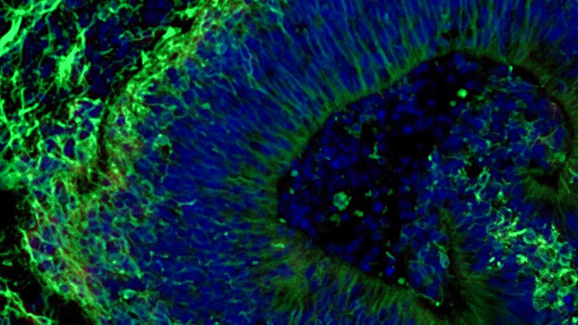
The motor cortex functions as the command center for initiating and controlling voluntary movement, exerting influence over the spinal motor neurons and, ultimately, muscle contractions. Its structural integrity is paramount for smooth, coordinated motor function. Using high-resolution diffusion MRI, the researchers quantitatively assessed microstructural properties of the motor cortex in a large cohort of Parkinson’s patients across a spectrum of severities. These microstructural features included metrics sensitive to neuronal density, dendritic complexity, and glial composition, collectively reflecting the tissue’s health and connectivity.
Intriguingly, the data revealed a consistent pattern of motor cortex microstructural degradation that correlated with motor symptom severity scores, independent of dopaminergic denervation markers derived from other imaging modalities. This dissociation suggests that pathogenic processes in the motor cortex itself, or upstream cortical circuits, contribute to symptom development in ways not captured by dopamine-centric models. The identification of these non-dopaminergic alterations represents a paradigm shift, emphasizing the significance of cortical involvement in Parkinson’s pathophysiology.
Furthermore, the study employed advanced statistical modeling to control for confounding factors such as age, disease duration, medication status, and global brain atrophy, underscoring the robustness of the association between motor cortex microstructure and clinical severity. These findings imply that neurodegeneration in Parkinson’s is a multi-region, multi-mechanistic process, with cortical microstructural changes serving as an independent biomarker of disease progression. This insight could transform diagnostic practices, enabling earlier identification of patients at risk for rapid motor decline.
On a mechanistic level, the observed microstructural changes may reflect synaptic loss, reduced dendritic arborization, or altered glial function within the motor cortex. Such alterations could impair intracortical connectivity and disrupt the fine-tuning of motor commands, exacerbating movement deficits. The study speculates that these cortical changes might be mediated by pathological protein aggregates or neuroinflammation intrinsic to Parkinson’s disease pathology, inviting further molecular investigations.
The implications for treatment are profound. Current dopaminergic therapies only partially alleviate motor symptoms and lose effectiveness over time, underscoring the need for alternative strategies. Targeting the motor cortex directly through neuromodulation techniques such as transcranial magnetic stimulation (TMS) or transcranial direct current stimulation (tDCS) might offer novel routes to restore cortical function. Moreover, neuroprotective agents focusing on cortical neurons and glial cells could emerge as complementary interventions to slow or halt disease progression.
This study also paves the way for more personalized medicine approaches in Parkinson’s. By quantifying motor cortex microstructural integrity non-invasively, clinicians could better tailor therapies and monitor responses beyond dopamine replacement efficacy. Longitudinal imaging could track disease trajectory with unprecedented precision, guiding timely therapeutic adjustments and prognostic counseling. Such refined patient stratification holds promise for improving quality of life and functional independence.
As technology advances, combining multimodal imaging techniques that integrate dopamine-specific PET scans with high-resolution MRI of cortical microstructure could yield comprehensive maps of Parkinson’s pathology. This holistic view will enhance our understanding of how cortical and subcortical changes interrelate and jointly influence motor and non-motor symptoms. Ultimately, unraveling these complex neural substrates may be key to developing holistic, disease-modifying treatments.
The study’s methodology deserves special mention. Employing cutting-edge diffusion MRI sequences and sophisticated analytic pipelines allowed the researchers to detect microstructural alterations at a voxel level previously unattainable in clinical populations. This technical rigor sets new standards for neuroimaging studies in neurodegeneration, highlighting the importance of continuous innovation in imaging technologies for neurobiological discovery.
Importantly, the authors acknowledge limitations, including the cross-sectional design which precludes causal inference, and the need for replication in diverse populations. Longitudinal studies will be critical to determine whether cortical microstructural changes precede or follow dopaminergic neuron loss and if they predict future motor decline. Additionally, integrating behavioral and electrophysiological data could enrich interpretations of cortical dysfunction.
The research invites a fundamental reconsideration of Parkinson’s disease as more than a dopamine disorder. It foregrounds the cortex’s vital role not only as a downstream target but as an active participant in disease processes. This expanded view compels reexamination of disease models, emphasizing circuit-level dysfunction encompassing cortical-subcortical pathways rather than isolated nigrostriatal degeneration.
In conclusion, the study by Honhar, Tinaz, Ebrahimian Sadabad, and colleagues heralds a new era in Parkinson’s research—one wherein cortical microstructure emerges as a key biomarker and potential therapeutic target. As we uncover the multifaceted nature of this devastating disorder, integrating cortical insights promises to enhance diagnosis, treatment, and ultimately, patient outcomes. The dopamine-independent association between motor cortex microstructure and disease severity opens thrilling possibilities for scientific exploration and clinical innovation in Parkinson’s disease.
Subject of Research: Parkinson’s disease pathology and motor cortex microstructure
Article Title: Neuroimaging evidence for a dopamine-independent association between motor cortex microstructure and Parkinson’s disease severity
Article References:
Honhar, P., Tinaz, S., Ebrahimian Sadabad, F. et al. Neuroimaging evidence for a dopamine-independent association between motor cortex microstructure and Parkinson’s disease severity. npj Parkinsons Dis. (2026). https://doi.org/10.1038/s41531-026-01411-x
Image Credits: AI Generated
Tags: advanced Parkinson’s diagnosticscortical contributions to Parkinson’s diseasedopamine-independent Parkinson’s mechanismsmicrostructural brain alterations in neurodegenerationmotor cortex microstructure in Parkinson’smotor cortex neuroarchitecture changesmotor symptoms variability in Parkinson’sneurodegeneration beyond dopamineneuroimaging in Parkinson’s researchnon-dopaminergic pathways in Parkinson’snovel therapeutic targets for Parkinson’sParkinson’s disease severity biomarkers


 English (US) ·
  French (CA) ·