

 2 hours ago
4
2 hours ago
4
PROTECT YOUR DNA WITH QUANTUM TECHNOLOGY
Orgo-Life the new way to the future Advertising by AdpathwayTiny Nanoparticles Could Transform the Fight Against Deadly Ventricular Arrhythmias
Ventricular fibrillation and other ventricular arrhythmias can turn a heartbeat into a medical emergency within seconds. In ventricular fibrillation, the heart’s lower chambers quiver chaotically instead of contracting, stopping effective blood flow and making sudden cardiac death a constant threat. Standard treatments, including antiarrhythmic drugs, catheter ablation and implantable cardioverter-defibrillators, can be lifesaving, but they do not fully solve the underlying problem. Drugs may affect tissues throughout the body, implanted devices cannot eliminate every arrhythmogenic substrate, and diagnosis may arrive only after dangerous electrical instability has already developed. A review by researchers in China now proposes that multimodal nanotechnology could eventually bring imaging, drug delivery and biological or physical therapy together in a single precision platform—while emphasizing that the concept remains far from routine clinical use.
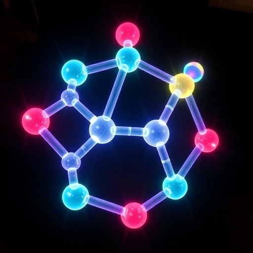
The proposed technology belongs to the emerging field of “theranostics,” a term combining therapy and diagnostics. Instead of acting as a passive drug container, a multifunctional nanoparticle could be designed to locate damaged or electrically unstable cardiac tissue, reveal it through imaging, release treatment in response to local biological signals and allow clinicians to monitor the result. Nanoparticles can be engineered with unusually large surface areas relative to their volume, adjustable sizes and shapes, and chemically modifiable surfaces. These properties allow scientists to attach targeting ligands, load several therapeutic compounds or incorporate imaging agents. Potential platforms include lipid carriers such as liposomes, polymeric nanoparticles and inorganic structures made from materials including gold, iron oxide or mesoporous silica. In principle, one particle could carry a magnetic-resonance contrast agent alongside an anti-inflammatory or anti-fibrotic drug, creating a molecular map of the heart while treating the tissue that generates abnormal electrical activity.
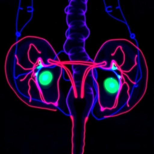
The biological target is not simply an irregular heartbeat but the complex tissue landscape that permits it. After a heart attack or chronic disease, ventricular muscle may undergo fibrosis, inflammation, altered ion-channel activity and disruption of connexin 43, a protein that helps electrical signals pass between neighboring cells. Scar tissue can form channels through which abnormal impulses circulate, while changes in autonomic nerves can further increase instability. The review describes possible molecular targets associated with these processes, including angiotensin II type 1 receptors in infarct scars, vascular cell adhesion molecule-1 on activated blood vessels, exposed myosin fragments in injured cardiomyocytes and tenascin-C in fibrotic tissue. Nanoprobes directed toward such markers could, in theory, identify dangerous regions more precisely than anatomical imaging alone. Magnetic resonance imaging could define scar structure, while photoacoustic imaging could provide information linked to light absorption by blood, lipids or engineered contrast materials.
The most important warning in the review is that the heart is not a tumor. Much of the inspiration for multimodal nanomedicine comes from oncology, where nanoparticles can sometimes exploit the enhanced permeability and retention effect. Tumors often have abnormal, leaky blood vessels that allow particles to escape into diseased tissue and remain there. The myocardial capillaries, by contrast, are continuous and have tight junctions reported to be roughly 4 nanometers wide. This makes passive entry of larger nanoparticles extremely limited, even in damaged or infarcted myocardium. Rapid coronary blood flow may also wash particles away, while the liver, spleen and other components of the reticuloendothelial system can remove them from circulation within minutes or hours. Cardiac nanomedicine therefore would need active targeting, such as receptor-mediated transport across endothelial cells, followed by retention or internalization in the myocardium. That strategy introduces new problems, including receptor saturation, competition with other molecules and substantial differences between patients.
The review highlights “smart” nanocarriers as one possible way to improve specificity. These particles could be programmed to open or change behavior in response to the chemical environment of diseased tissue. Acidosis associated with ischemia, oxidative stress and increased matrix-metalloproteinase activity during remodeling are among the triggers that might be exploited. Mesoporous silica nanoparticles, for example, have a rigid, high-surface-area framework capable of holding substantial amounts of drug. Molecular caps could be designed to detach under a particular pH or after exposure to an enzyme, releasing the payload only after the particle reaches a pathological compartment. Biomimetic coatings may offer another solution. Nanoparticles cloaked in membranes derived from cardiac cells could inherit surface proteins that improve recognition of cardiac tissue while reducing uptake by macrophages. Such approaches remain experimental, and a coating that improves delivery in one type of scar or inflammatory environment may perform poorly in another.
Physical therapies could also be integrated into the nanoscale systems. In photothermal therapy, materials such as gold nanorods or platinum nanoshells absorb near-infrared light and convert it into heat. In photodynamic therapy, light activates a compound that generates reactive oxygen species, molecules capable of damaging selected cells. The goal in arrhythmia treatment would not necessarily be to destroy large regions of the heart, but to modulate a critical scar channel, influence autonomic nerves or alter a localized electrophysiological circuit. A cited canine study used platinum nanoshells with a reported photothermal conversion efficiency of 73.7 percent at a wavelength in the near-infrared-II range. By controlling temperature in the left stellate ganglion, researchers activated TRPV1 channels at approximately 41.0–42.9 degrees Celsius and inhibited activity through TREK1 channels at about 45.0–46.9 degrees Celsius. This bidirectional autonomic modulation reduced arrhythmias associated with ischemia and reperfusion, with no detectable neuronal damage or systemic toxicity during a 30-day follow-up.
Other preclinical findings point toward immune and direct electrical mechanisms. An oxygen-independent photodynamic nanocomposite was reported to selectively reduce pro-inflammatory M1 macrophages in the left stellate ganglion after myocardial infarction in beagles. Conventional photodynamic therapy depends on oxygen, a serious limitation in ischemic tissue, but oxygen-independent designs could potentially work under hypoxic conditions. Separately, red-light-activated conjugated polymers altered the behavior of human stem-cell-derived cardiomyocytes without genetic modification. The treatment accelerated calcium cycling, shortened action potentials and increased spontaneous beating frequency through pathways involving the sarcoplasmic reticulum calcium ATPase and the sodium-calcium exchanger. These studies offer striking proof-of-concept results, but they do not establish a treatment for people. Light may penetrate only about 1–2 centimeters through tissue in the near-infrared-I range and 2–3 centimeters in the near-infrared-II range, leaving deep ventricular or septal targets difficult to reach. The heart’s constant movement adds another layer of complexity.
Safety may be the greatest barrier. A nanoparticle intended to stabilize electrical activity could theoretically create a new arrhythmogenic substrate by interfering with ion channels, increasing oxidative stress or altering inflammatory signaling at gap junctions. Reactive oxygen species, useful at carefully controlled doses, can damage lipids, mitochondria, proteins and DNA when generated in excess. Heat that spreads beyond a target could injure viable myocardium or create new conduction barriers. Long-term retention is another concern because the heart has limited regenerative capacity and may be vulnerable to cumulative exposure. Researchers would need to track particle distribution, degradation products, liver and kidney effects, blood counts, coronary integrity and electrical function over extended periods. The review also points to practical hurdles: complex particles are difficult to manufacture reproducibly, combination products that function as both diagnostic agents and therapies do not fit neatly into existing regulatory categories, and the appropriate dose may vary with the patient’s scar structure, perfusion and immune status.
The proposed technological leap would therefore require a parallel transformation in clinical care. Nurses would be central to any future implementation, not merely to routine observation but to managing the new risks created by nanomedicine. Before administration, teams would need to assess baseline ECG findings, cardiac function, kidney and liver performance, bleeding risk, allergies and possible interactions with existing medicines. During infusion, staff would watch for complement activation-related pseudoallergy, a reaction that can cause flushing, breathlessness, low blood pressure or chest pain. After treatment, continuous ECG surveillance could focus on QT-interval changes, premature ventricular complexes, non-sustained or sustained ventricular tachycardia and altered heart-rate variability. Standardized protocols would also specify infusion rates, observation periods, laboratory testing, adverse-event reporting and criteria for stopping treatment. Electronic health records, telemetry systems, wearable patches and machine-learning tools could help detect deterioration, but only if staff are trained to interpret their warnings and recognize artifacts.
The review ultimately presents multimodal nanotechnology as a roadmap rather than a ready-made therapy. Future research will need cardiac-specific targeting strategies, testing in human induced pluripotent stem-cell-derived cardiomyocytes, ex vivo perfused hearts and large-animal models, as well as long-term studies lasting at least a year. Researchers may eventually combine nanoparticles with implantable or bioresorbable bioelectrodes that map electrical activity and deliver a calibrated response in a closed loop. Artificial intelligence could help integrate imaging, genomic, proteomic and metabolic data to predict which substrate is most likely to trigger an arrhythmia and select the appropriate payload. Yet the central promise will be meaningful only if it survives rigorous safety testing, scalable manufacturing and clinical trials. For now, the most credible breakthrough is conceptual: a future in which the same nanoscale system can see the dangerous tissue, reach it despite the heart’s formidable vascular barriers, treat it without destabilizing healthy myocardium and guide clinicians through every step of care.
Subject of Research: Multimodal nanotechnology and standardized nursing management for ventricular arrhythmias
Subject of Research: Medicine
Article Title: Application of multimodal nanotechnology and standardized nursing management in ventricular arrhythmia
Article References: Ding, X., Zhang, L., Guo, F., Pan, X., Wei, X., Su, L., Wu, L., Min, L., Zhang, M., & Han, L. (2026). Application of multimodal nanotechnology and standardized nursing management in ventricular arrhythmia. BioMedical Engineering OnLine, 25(1), Article 77. https://doi.org/10.1186/s12938-026-01571-0
Image Credits: AI Generated
DOI: 10.1186/s12938-026-01571-0
Keywords: ventricular arrhythmia, multimodal nanotechnology, theranostics, precision medicine, targeted drug delivery, photothermal therapy, nursing management, cardiac safety
Cite this news
APA MLA Chicago
SCIENMAG. (August 28, 2026). Multimodal Nanotechnology and Standardized Nursing Management Applied to Ventricular Arrhythmia. https://scienmag.com/multimodal-nanotechnology-and-standardized-nursing-management-applied-to-ventricular-arrhythmia/
Copy citation Download RIS
Tags: advanced cardiac tissue imaging techniquesadvanced nanoparticle platforms for arrhythmia managementbiological signal-responsive nanoparticlesclinical prospects of multimodal nanomedicine in arremerging nanotech applications in cardiologyemerging nanotech solutions for arrimaging and therapy integration in cardiologyimaging-guided cardiac therapyintegrated therapy and diagnostics for heart rhythm disordersmultimodal nanomedicine in cardiac therapymultimodal theranostics for cardiac arrhythmiasnanoscale biological sensors for arrhythmia detectionNanotechnology for ventricular arrhythmia treatmentnanotechnology in arrhythmia managementnanotechnology-based cardiac tissue repairprecision nanomedicine in cardiac careprecision nanomedicine in cardiologystandardization in nanomedicine for heart diseasesstandardized nursing management in ventricular arrhythmia treatmenttargeted drug delivery for ventricular fibrillationtargeted nanoparticle drug delivery for ventricular fibrillationtheranostic nanoparticles for heart arrhythmiasVentricular arrhythmia nanotechnology
















 English (US) ·
  French (CA) ·