

 6 hours ago
15
PROTECT YOUR DNA WITH QUANTUM TECHNOLOGY
Orgo-Life the new way to the future Advertising by AdpathwayNeurotrophin-3 (NT-3) bidirectionally regulates oligodendrocyte autophagy after spinal cord injury, a process mediated by a temporal switch from the P75NTR to the TrkC receptor. In a rat model of spinal cord injury, neurotrophin-3, a growth factor naturally concentrated in spinal cord tissue, first pushed oligodendrocyte autophagy upward during the acute phase and then pulled it back down during the subacute phase. That reversal was not a random fluctuation. The researchers traced it to a receptor switch: NT-3 initially signaled through P75NTR to activate AMPK and restrain mTOR, and later through TrkC to activate the PI3K/AKT/mTOR axis. The pivot between the two was driven by ubiquitination-dependent endocytosis and degradation of P75NTR, which freed NT-3 to bind its second receptor.
The study, published in Frontiers in Molecular Neuroscience, combined a rat compression model of spinal cord injury with an oxygen-glucose deprivation model in primary oligodendrocytes. Eighty-four eight-week-old Sprague-Dawley rats were used, with spinal cord compression applied for ten seconds via a vascular clip after laminectomy at the T10-T11 level. Motor recovery was scored on the Basso-Beattie-Bresnahan locomotor scale by two blinded investigators at days 1, 3, 7, and 14. In the treated animals, NT-3 was delivered intrathecally through a catheter placed at the L3/L4 intervertebral space, at a dose of 20 micrograms per kilogram daily for seven consecutive days.
The functional results were clear-cut. Sham-operated rats kept maximum BBB scores of 21 throughout. Injured rats without treatment scored a median of 1.5 on day 1, 2.0 on day 3, 4.0 on day 7, and 15 on day 14. With NT-3, the medians rose to 2.0, 6.5, 8.0, and 16 at the same time points. Dunn’s post-hoc analysis confirmed the treated group scored significantly higher than the untreated injured group at days 3, 7, and 14, though not on day 1. Endogenous NT-3 itself rose progressively at days 1, 3, and 7 after injury, which the authors interpret as part of the body’s own repair response.
Protection of oligodendrocytes, the myelin-forming cells of the central nervous system, emerged as the cellular basis of that recovery. At day 7, double immunofluorescence for CD68 and myelin basic protein showed far more myelin debris inside CD68-positive phagocytes in untreated injured rats than in either sham or NT-3-treated animals. Because MBP labels both intact myelin and debris, debris within CD68 cells served as the readout of oligodendrocyte injury. Treated animals also showed significantly higher expression of proteolipid protein 1, a marker of mature oligodendrocytes, and histopathology revealed abundant oligodendrocytes surrounding axons in the treated group, while the untreated injured group had no intact axonal structures in white matter and only scattered oligodendrocytes.
The autophagy story unfolded in two acts. Western blotting showed autophagic activity, measured through the LC3BII-to-LC3BI ratio and the autophagy substrate P62, rising progressively in untreated injured spinal cord. In NT-3-treated animals the response was biphasic: elevated at day 3, then markedly reduced at day 7. The same temporal pattern appeared in cultured oligodendrocytes deprived of oxygen and glucose, where autophagy climbed by 3 hours after NT-3 treatment and fell to low levels by 12 hours. To test whether this mattered for survival, the team used 3-methyladenine to block autophagy and rapamycin to force it. Either manipulation alone triggered substantial oligodendrocyte apoptosis, supporting the idea that phase-tuned autophagy, not simply more or less of it, protects the cells.
Receptor knockdown experiments pinned each phase to a specific receptor. Silencing P75NTR at the 3-hour mark abolished the NT-3-driven rise in autophagy, while silencing TrkC at 12 hours prevented the later suppression, leaving autophagic activity persistently high. Downstream, phosphorylation analysis showed AMPK activation with mTOR inhibition at 3 hours, and PI3K/AKT/mTOR activation at 12 hours. Because AMPK promotes autophagy and PI3K/AKT signaling suppresses it, the two pathways neatly explain the flip from promotion to inhibition.
A re-analysis of public translatome data added independent, if indirect, support. The GSE225308 dataset profiles HA-immunopurified mature oligodendrocyte translatomes from RiboTag mice after moderate T9 contusion, at naive, 2 days, and 10 days post-injury, with three biological replicates per group. Across 39 autophagy-lysosome genes, the program showed progressive upregulation after injury: lysosomal hydrolases and a V-ATPase subunit were already elevated at 2 days, the ULK1 initiation module was transiently downregulated at that stage, and by 10 days initiation had recovered while lysosomal genes stayed high. The authors caution that autophagic flux is governed by post-translational events, so gene expression cannot reflect true flux; the analysis serves as contextual evidence only.
Correlation analysis across the nine mature oligodendrocyte samples linked the two receptors to opposite tendencies. Ngfr, the gene encoding P75NTR, correlated positively with Map1lc3b, Gabarapl1, Sqstm1, Optn, Ctss, Tfeb, and Hexb, with r values from 0.69 to 0.87 and nominal p values below 0.05. Ntrk3, encoding TrkC, correlated negatively with Mitf, a regulator of lysosomal biogenesis, at r = -0.84. Ntf3 itself correlated negatively with Atg3. These are associations in a small sample, not causal demonstrations, but they align with the mechanistic experiments.
The receptor switch itself was the most striking finding. In spinal cord tissue at the subacute phase, double staining for P75NTR and EEA1, a marker of early endosomes, showed significant internalization of P75NTR in treated animals, quantified by Pearson correlation. Co-immunoprecipitation detected higher levels of ubiquitinated P75NTR in the treated group. In vitro, the endocytosis inhibitor cytochalasin B and the ubiquitination inhibitor PYR41 each blocked the formation of endocytic vesicles and kept P75NTR levels high at both 3 and 12 hours, whereas untreated NT-3-treated cells showed P75NTR elevated at 3 hours but back to baseline by 12 hours. Transfecting cells with tagged ubiquitin and P75NTR plasmids confirmed increased ubiquitinated P75NTR after NT-3, an effect abolished by UBE1 siRNA. Reverse co-immunoprecipitation on spinal cord lysates showed that NT-3 binding to TrkC was not significantly elevated at day 3 but was significantly enhanced at day 7, consistent with TrkC becoming the predominant receptor in the subacute phase.
The authors are careful about limits. The in vivo P75NTR internalization was not directly localized to oligodendrocytes, the in vitro setting cannot fully reproduce the injured cord’s multicellular environment, and no in vivo knockdown experiments tested autophagy regulation by each receptor at the two stages. Sample sizes for Western blotting and immunofluorescence were limited, and the specific mechanism inducing P75NTR ubiquitination remains unresolved. NT-3 also acts on neurons, astrocytes, microglia, and progenitor cells, so off-target effects are possible, and the safety of intrathecal delivery in terms of dose, duration, and spread requires preclinical and early-phase clinical evaluation. What the study does establish, within those bounds, is a mechanistic chain in a rodent model: a growth factor that times its own receptor traffic to first clear damaged components and then pull the brakes on autophagy, keeping myelin-forming cells alive long enough to repair.
Subject of Research: NT-3 receptor switching and bidirectional autophagy regulation in oligodendrocytes after spinal cord injury
Article Title: NT-3 promotes functional recovery after spinal cord injury by bidirectionally regulating oligodendrocyte autophagy via P75NTR/TrkC receptor switching
Article References: Cong, Y., Wang, P., Sun, Z., & Yu, J. (2026). NT-3 promotes functional recovery after spinal cord injury by bidirectionally regulating oligodendrocyte autophagy via P75NTR/TrkC receptor switching. Frontiers in Molecular Neuroscience, 19, Article 1951547. https://doi.org/10.3389/fnmol.2026.1951547
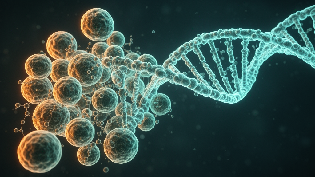
Image Credits: AI Generated
DOI: 10.3389/fnmol.2026.1951547
Keywords: spinal cord injury, neurotrophin-3, oligodendrocytes, autophagy, P75NTR, TrkC, receptor switching, ubiquitination, endocytosis, myelin repair, BBB score, rat model


















 English (US) ·
 French (CA) ·