

 6 hours ago
11
6 hours ago
11
PROTECT YOUR DNA WITH QUANTUM TECHNOLOGY
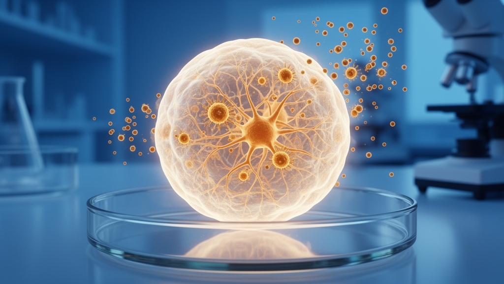
Orgo-Life the new way to the future Advertising by AdpathwayFor more than half a century, developmental biologists have chased a deceptively simple question: what is the minimum set of instructions needed to launch a mammalian embryo? A fertilized egg clearly carries the full program, but whether a single, non-germline cell pushed into an embryonic state could autonomously rebuild an entire organism has remained stubbornly out of reach. A team led by Sheng Ding at Tsinghua University now reports in Nature that it has come closer than ever to that goal. Starting exclusively from chemically induced totipotent-like stem cells, or ciTotiSCs, the researchers coaxed self-organizing structures through the morula and blastocyst stages, past implantation and gastrulation, and all the way to early organogenesis, with extended cultures producing functional cardiac tissue comparable to that of a natural ten-and-a-half-day-old mouse embryo.
The starting material is the product of a chemical strategy the same group pioneered in 2023. Rather than introducing transcription factors or relying on rare, transient subpopulations of embryonic stem cells, the team used a defined cocktail of small molecules to push mouse cells into a state that molecularly mirrors the two-cell embryo, the earliest stage at which the zygotic genome has just switched on. Single-cell RNA sequencing of the resulting ciTotiSCs revealed strong activation of both maternal and zygotic genome activation programs, hallmarks of genuine totipotency, alongside suppression of the pluripotency networks that define conventional embryonic stem cells. In other words, the cells were not merely pluripotent, capable of making embryonic tissues, but appeared to carry the broader potential that includes the extraembryonic lineages needed to build a placenta and yolk sac.
Converting that molecular resemblance into actual embryogenesis, however, demanded a carefully choreographed culture environment. The researchers aggregated ciTotiSCs in microwells and exposed them to a sequence of media conditions, including combinations of small molecules and growth factors such as A83-01, CHIR, LPA, EGF and BMP4, followed by heparin and FGF4. Within days, the aggregates formed blastocyst-like structures that closely resembled natural blastocysts in both morphology and lineage segregation. Immunostaining showed the expected spatial organization: SOX2- and OCT4-positive cells forming the inner cell mass, GATA3-positive cells forming the trophectoderm, and GATA6- and SOX17-positive cells marking the primitive endoderm. Even the squamous, stretched morphology of natural trophectoderm cells was reproduced, and the models displayed rhythmic cycles of blastocoel cavity inflation and deflation reminiscent of living blastocysts.
What followed is arguably the most striking part of the study. Under conditions supporting continuous ex utero development, the ciTotiSC-derived embryo models, or ciToti-EMs, progressed through peri-implantation stages, forming egg cylinder-shaped structures with a pro-amniotic cavity enclosed by SOX2-positive epiblast cells showing apical F-actin and ZO-1 enrichment, precisely as in natural embryos. The models then segregated their embryonic and extraembryonic compartments, developed visceral endoderm and parietal endoderm, and initiated gastrulation, marked by the appearance of Brachyury- and FOXA2-positive cells at a primitive streak. By the equivalent of embryonic day 8.5, the models displayed an anterior-posterior axis, neural folds expressing PAX6, somites marked by HOXB4, a hindgut, and regional specification along both the anterior-posterior and dorsal-ventral axes.
The extraembryonic lineages, often the weak point of synthetic embryo models, were also faithfully generated. Single-cell transcriptomics identified amnion, allantois, yolk sac and parietal endoderm populations in the day-8.5-equivalent models, and Pearson correlation analysis showed transcriptome-wide similarity between these compartments and their natural counterparts. Primordial germ cells emerged as well, identified by co-expression of BLIMP1 and STELLA, and could be tracked migrating through the tail bud and hindgut regions of the models. When dissociated and expanded on feeder layers, these cells showed the epigenetic reprogramming characteristic of the germline, including reduced H3K9me2 and elevated H3K27me3, and progressed toward maturation markers such as DAZL and DDX4. This suggests the ciToti-EM system could become a scalable source of germline cells for studying fertility and heredity.
Blood formation, another milestone of mid-gestation development, proceeded with remarkable fidelity. The models generated vascular endothelium, erythroblasts, hematopoietic epithelia and macrophage populations that clustered alongside their natural E8.5 equivalents in integrated single-cell analyses. Erythroblasts collected from the models were nucleated, as expected for primitive mouse blood, and colony-forming unit assays confirmed that the models contained functional hematopoietic progenitors capable of generating erythroid and macrophage colonies in vitro. The supervised comparison framework the team built, which trains a logistic regression classifier on an annotated atlas of natural embryo data and scores how well each model’s cell types match reference lineages, placed the ciToti-EMs ahead of previously published embryo models across most lineages and stages examined.
Perhaps the most visually compelling result came from extended culture. The ciToti-EMs developed beating heart-like structures, and live imaging captured peristaltic-like contractions in earlier models followed by coordinated, chamber-specific rhythmic beating in later ones. Histological and immunofluorescent analyses of the cardiac tissue revealed a thin epicardial layer, trabeculated myocardium, and regionalized expression of atrial, ventricular and atrioventricular canal markers, closely mirroring natural E10.5 hearts. Single-cell profiling distinguished first heart field, second heart field, atrial and ventricular cardiomyocytes, outflow tract cells, cardiac neural crest cells and endothelial cells, and even identified macrophages and erythroblasts within the cardiac chambers, as seen in natural embryos at this stage.
The implications reach well beyond mouse developmental biology. A model built entirely from a single, chemically induced cell type sidesteps the ethical and practical constraints of using natural embryos or mixtures of multiple stem cell lines, and offers a reproducible, scalable platform for dissecting the molecular logic of early mammalian development. Because the entire process begins with small molecules rather than genetic manipulation, it also hints at a future in which multicellular organisms, or at least substantial portions of them, could be generated from ordinary somatic cells without ever passing through a fertilized egg. The authors are careful to frame the work as a model system, and the models, while extraordinarily complete, are not viable organisms. Nonetheless, the demonstration that one induced cell type can autonomously recapitulate the full spectrum of early mouse embryogenesis marks a watershed for synthetic embryology.
Significant questions remain. The efficiency of progressing from starting aggregates to late-stage models is still modest, and the extrapolated developmental efficiencies suggest substantial attrition at each transition. Whether the same chemical strategy can be adapted to human cells, and what regulatory frameworks should govern such models, are debates that will intensify. For now, the ciToti-EM system gives researchers something they have long lacked: a self-contained, tractable window into mammalian development from the two-cell state to the beating heart, one that can be perturbed, imaged and sequenced at every step. As embryo models grow ever more faithful, the line between modeling life and initiating it grows thinner, and this study moves that line decisively forward.
Subject of Research: Modelling mouse embryogenesis from chemically induced totipotent stem cells
Article Title: Modelling mouse embryogenesis from chemically induced totipotent stem cells
Article References: Modelling mouse embryogenesis from chemically induced totipotent stem cells. (n.d.). https://doi.org/10.1038/s41586-026-11164-5
Image Credits: AI Generated
DOI: 10.1038/s41586-026-11164-5
Keywords: totipotent stem cells, embryo models, mouse embryogenesis, gastrulation, synthetic embryos, ciTotiSCs, blastocyst, cardiogenesis, hematopoiesis, primordial germ cells, single-cell RNA sequencing, developmental biology

















 English (US) ·
  French (CA) ·