

 1 week ago
13
1 week ago
13
PROTECT YOUR DNA WITH QUANTUM TECHNOLOGY
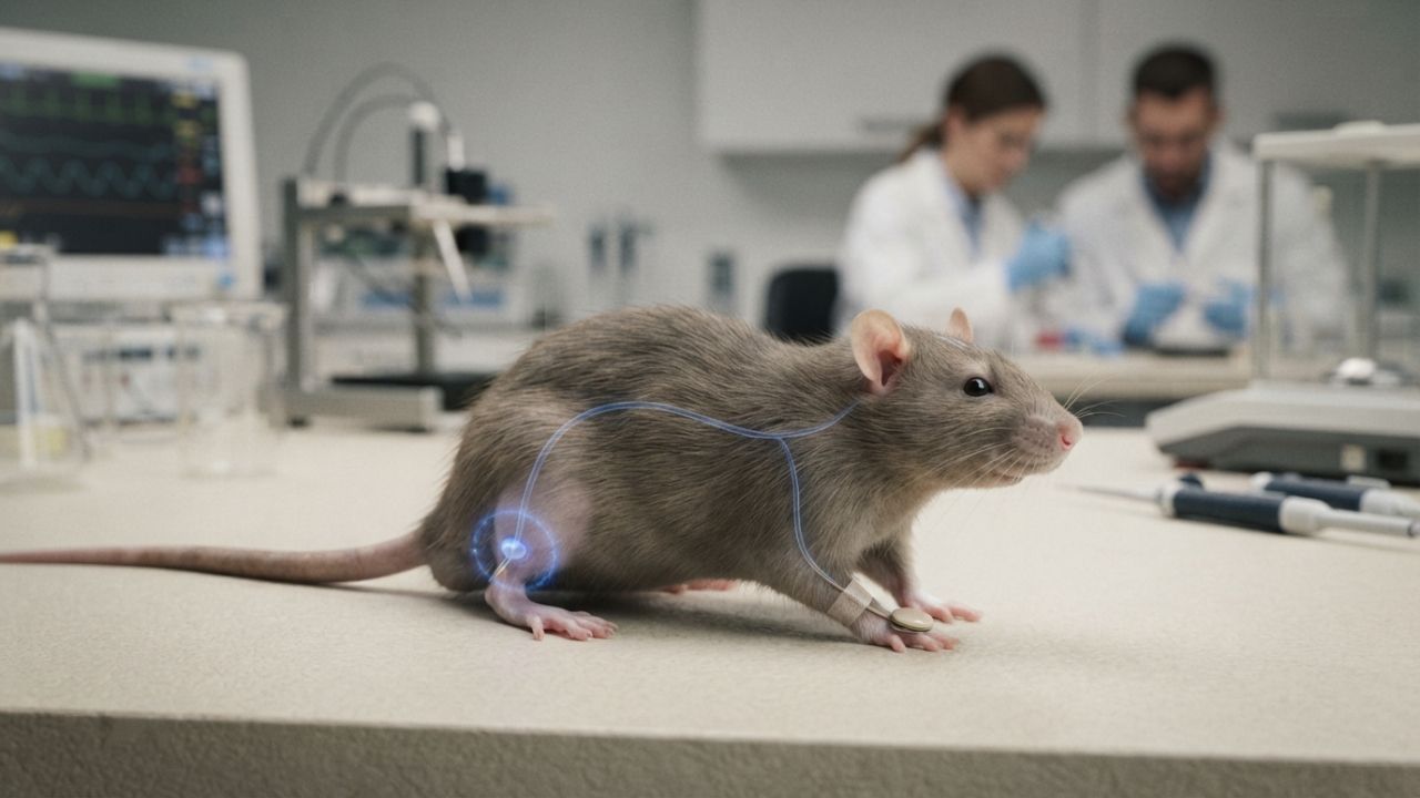
Orgo-Life the new way to the future Advertising by AdpathwayStanford Medicine researchers have grown human brain tissue inside mice that were engineered to develop almost none of their own cerebral cortex, and the transplanted cells produced a rare type of human neuron that scientists had never managed to create in laboratory culture. The study, published Sept. 16 in the journal Nature, reports that three months after surgery, more than 90% of the cortical tissue in the animals' brains by volume was human.
The researchers call the animals "xenocortical" mice, but they are not mice with human minds. Pasca has said that labels such as "humanized mice" or "mice with human brains" are inaccurate, because the animals retain a mouse nervous system. The human tissue remained developmentally immature, was not organized into the layers of a mature cortex and contained few inhibitory neurons. What the model offers is a living system for studying how human brain cells develop, connect and fail.
The work matters beyond the laboratory because it raises familiar questions about animal research, oversight and where scientists should draw lines. It also has an unexpected wildlife connection, since the rare neurons it produced are found in only a handful of large-brained animals, including whales, elephants and great apes.
Clearing Space for Human Tissue
Senior author Sergiu Pasca, a Stanford professor of psychiatry and behavioral sciences, had previously transplanted human cortical organoids, small lab-grown clusters of brain tissue made from stem cells, into newborn rats. The human cells developed far better inside a living brain, but the rat neurons matured much faster. "The two parallel developing systems are in competition for turf," Pasca said in Stanford Medicine's announcement.
The new study removed that competition. The team engineered "apallial" mice in which the cells that normally build most of the cortex and hippocampus never form, leaving adults with only about 2% of the usual cortical tissue. These mice survived in good health with only minor quirks, including a slightly more cautious gait and a tendency to forget recently encountered new environments.
Two days after birth, the mice received human cortical organoids, typically more than one per animal, each containing perhaps 100,000 cells from healthy donors who had consented to animal transplantation. Most of the surgeries succeeded. The human tissue expanded to fill the empty space and formed connections through the mouse brain and on to the spinal cord. As The Transmitter reported, graft neurons fired in large, synchronous bursts every few minutes, a sign that the tissue was forming a functional network.
A Rare Cell Type Shared with Whales, Elephants and Apes
The most surprising result was the appearance of cells the researchers identified as von Economo neurons, large, cigar-shaped cells that make up roughly one in every 90,000 cortical neurons. According to Stanford, they had previously been observed only in postmortem brain tissue and had never been produced in culture or in earlier, more crowded rodent transplants. The Nature paper describes the grafts as containing a distinct population of human projection neurons carrying molecular markers associated with these cells.
Once thought to be uniquely human, von Economo neurons have since been identified in other large-brained, highly social mammals, including great apes, elephants, dolphins and whales. In people, they sit in brain regions tied to social awareness and decision-making, and they appear especially vulnerable in some forms of frontotemporal dementia.
That gives researchers a new way to study the cells in a living, behaving animal. Pasca has said that while animal models have been extremely helpful, "some biological features seem to be uniquely human," which is why rodent brains alone have fallen short for studying many psychiatric and neurological conditions.
Behavior, Oxygen Deprivation and Disease Modeling
Stanford reported that three to six months after surgery, the xenocortical mice performed generally similarly to normal mice on behavioral tests. The Nature paper adds nuance, describing broadly preserved movement alongside selective differences in limb coordination and in how spontaneous behavior was organized. Outside experts have cautioned that the study does not show that human neurons are driving specific behaviors.
In a proof-of-concept test, the team exposed mice to five hours of low oxygen. The human tissue suffered substantial damage, and those mice had trouble keeping a steady gait and balance, reminiscent of cerebral palsy in children. Normal and apallial mice were virtually unaffected. Pasca said the model could eventually help researchers study schizophrenia, epilepsy, profound autism and cerebral palsy, and test possible treatments.
The evidence has clear limits. This is a single, peer-reviewed laboratory study in mice, not a treatment. The human tissue resembled developing cortex rather than a mature adult brain; it still matured on a slower human timeline, and the approach is limited by the short lifespan of mice. Joseph Gleeson, a neuroscientist at the University of California, San Diego, who was not involved, noted that variability in how the organoids grow and integrate could make some questions hard to answer.
Ethics, Oversight and a Primate Red Line
Placing human brain tissue into animals remains contentious. Some scientists and ethicists have raised concerns about introducing human abilities, or even consciousness, into animals. Ben Hurlbut, a biosciences ethicist at Arizona State University, told Science that such studies are "experimenting with concepts that are socially and morally meaningful."Madeline Lancaster, a brain organoid researcher at the Medical Research Council's Laboratory of Molecular Biology, told the journal that transplant work is ideally used to reveal what lab-dish models are missing so those models can improve.
Pasca has said he is not concerned that the mice gained human cognitive capacities, pointing to their tiny brains and the vast evolutionary gap between mice and people. He told MIT Technology Review that performing the same experiment in a primate would be "a very clear red line." Stanford said the team sought input over several years from ethicists, neurobiologists, patient advocates, philosophers and legal scholars, and Pasca organized a conference at Asilomar, California, in November 2025 to debate the ethics of human stem cell models and their transplantation.
Readers should also know about financial interests. Stanford's Office of Technology Licensing holds patents on generating cortical organoids with Pasca listed as an inventor, and a provisional patent application on organoid transplantation names Pasca and several co-authors. The work was funded by the Stanford Wu Tsai Neurosciences Institute, the Kwan Funds, the Senkut Funds and the Brain & Behavior Research Foundation.
For families living with autism, epilepsy or cerebral palsy, nothing about current care changes. Stanford said a drug candidate that emerged from the lab's earlier research on Timothy syndrome is being prepared for early-stage safety trials, and Pasca received a $14 million grant earlier this month to continue studying neurodevelopmental disorders. The central open question is whether findings in xenocortical mice will translate into treatments for people.
What Readers Want to Know
What are xenocortical mice?
They are mice engineered to develop almost none of their own cerebral cortex, then given transplants of lab-grown human cortical tissue two days after birth. The human tissue grew to make up more than 90% of their cortical tissue by volume.
Do these mice think like humans?
No. The human tissue stayed immature and lacked the layered structure of a mature cortex, the mice performed broadly like normal mice rather than better, and Pasca has said he does not believe the mice gained human cognitive abilities.
What are von Economo neurons?
They are rare, large neurons found in brain regions tied to social awareness. Besides humans, they occur in great apes, elephants, dolphins, and whales, and they have never been generated in a lab dish before.
Why would scientists do this research?
Living human brain tissue is almost impossible to study directly. The model lets researchers watch human brain cells develop and respond to injury, such as oxygen loss linked to cerebral palsy, inside a living animal.
Where was the study published?
It was published Sept. 16, 2026, in the journal Nature. Science, MIT Technology Review, and other outlets published news coverage of the findings.
Are there limits on this kind of research?
Stanford says the work followed years of ethics consultation, and Pasca has called transplanting human brain organoids into primates a clear red line. Critics say the research still raises unresolved moral questions.
Does this change treatment for autism or cerebral palsy?
Not now. It is an early laboratory model. Any treatments developed with its help would still need years of testing in clinical trials.
© 2026 NatureWorldNews.com All rights reserved. Do not reproduce without permission.




















 English (US) ·
  French (CA) ·