

 5 hours ago
8
PROTECT YOUR DNA WITH QUANTUM TECHNOLOGY
Orgo-Life the new way to the future Advertising by AdpathwayCognitive impairment represents a major and persistent determinant of poor functional recovery in schizophrenia, yet the exact molecular events connecting neuroimmune disturbance, cellular redox imbalance, and synaptic dysfunction remain incompletely understood. In a narrative mechanistic review published in Frontiers in Psychiatry, researchers led by Fang Liu from the Department of Psychiatry at The First Affiliated Hospital of Kunming Medical University synthesized current clinical and experimental literature to propose a testable biological hypothesis. The authors evaluated how circulating extracellular vesicle microRNAs, nitric oxide synthase and nitric oxide regulation, and NLR family pyrin domain containing 3 inflammasome signaling might converge to influence synaptic injury and cognitive phenotypes. Rather than claiming a confirmed pathogenic sequence, the authors emphasized the evidentiary boundaries separating preliminary molecular associations from established causal mechanisms, highlighting that altered profiles across separate biological tiers do not yet prove a continuous pathogenic pathway in human patients.
The review established that cognitive deficits in schizophrenia emerge early during neurodevelopment and span multiple operational domains, including attention, working memory, learning, processing speed, executive function, and social cognition. While standard antipsychotic medications can alleviate positive psychiatric symptoms, their effectiveness in improving cognitive outcomes remains markedly limited. The authors outlined a multifaceted pathological network wherein genetic susceptibility interacts with environmental insults, prenatal infections, and perinatal complications to disrupt synapse formation, brain network maturation, and neurotransmitter balance across dopaminergic, glutamatergic, and GABAergic systems. Within this framework, neuroinflammation has emerged as a critical bridge linking underlying molecular disturbances to cognitive deterioration. Clinical investigations have frequently recorded elevated peripheral inflammatory markers such as interleukin-1 beta, interleukin-6, tumor necrosis factor-alpha, and C-reactive protein in patients, although these observational correlations vary considerably depending on illness duration, metabolic status, antipsychotic exposure, and detection methodology.
Central nervous system glial cells, particularly microglia, play a more direct role in maintaining cognition-related neural circuits than general peripheral inflammatory markers. Under normal physiological conditions, microglia monitor the neural microenvironment, modulate synaptic plasticity, and eliminate weak or redundant synapses through complement-dependent mechanisms to support circuit maturation. However, under conditions of persistent immune stimulation, genetic risk, or oxidative stress, microglia can shift into aberrant activation states that release pro-inflammatory cytokines such as interleukin-1 beta and interleukin-18, generate reactive oxygen species, and promote excessive synaptic elimination. Genetic discoveries linking structural variations in the complement component C4 gene to schizophrenia risk, alongside patient-derived cellular models demonstrating enhanced synaptic phagocytosis and diminished synaptic density, indicate that excessive complement tagging and aberrant microglial pruning contribute directly to functional connectivity loss. Nevertheless, the authors observed that peripheral immune shifts alone remain heterogeneous and insufficient to define precise intervention targets, underscoring the necessity of identifying upstream regulatory drivers.
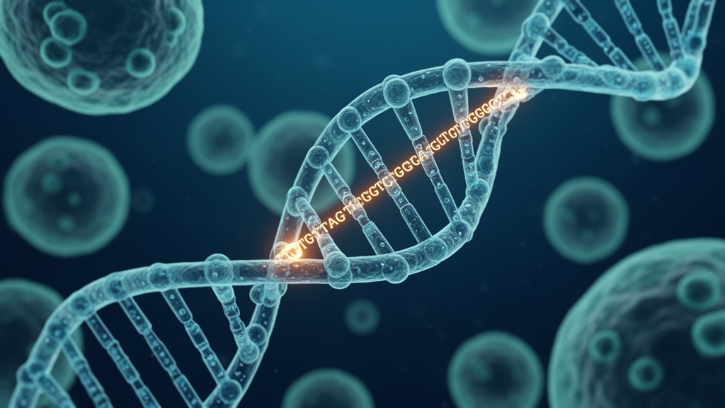
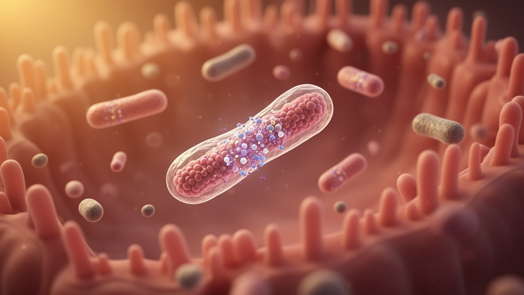
Extracellular vesicles and their microRNA cargo have emerged as key candidates for mediating intercellular communication and multi-target transcriptional regulation between the peripheral circulation and the central nervous system. Following the Minimal Information for Studies of Extracellular Vesicles recommendations, the authors noted that lipid bilayer-enclosed extracellular vesicles transport bioactive proteins, lipids, and nucleic acids across biological barriers, including the blood-brain barrier via transcytosis. A critical examination of existing patient cohorts demonstrated that while differential microRNA expression patterns occur in circulating vesicles, reported signatures vary widely across studies. For instance, Barnett and colleagues isolated microtubule-associated protein 1B-enriched serum vesicles of putative neuronal origin from 221 patients with schizophrenia and 256 controls, finding distinct microRNA profiles in patients with cognitive impairment whose predicted targets clustered in neurodevelopmental and postsynaptic pathways. However, most participants had longstanding illness, 208 were receiving antipsychotics, and 32 of the 42 clozapine-treated patients were in the cognitive-deficit subgroup, complicating the separation of cognitive subtypes from medication effects and treatment resistance.
Methodological discrepancies across clinical studies further constrain direct comparisons of extracellular vesicle microRNA profiles. Chen and colleagues examined small plasma vesicles isolated by size-exclusion chromatography in a screening cohort of 24 individuals followed by validation in 89 patients and 50 controls, reporting elevations in miR-23a, miR-103a, and miR-450b that were not mirrored in vesicle-depleted plasma. Tomita and colleagues utilized chemical precipitation followed by L1 cell adhesion molecule immunocapture in 14 recent-onset patients and eight chronic-phase patients alongside matched controls, detecting reduced miR-184 at both illness stages. The authors cautioned that L1CAM specificity remains debated because circulating L1CAM often exists as a soluble free protein, while small, unequal cohorts and substantial differences in chlorpromazine-equivalent medication doses between recent-onset and chronic patients hinder stage-specific conclusions. Studies in first-episode psychosis by Xavier and colleagues observed reductions in miR-335-5p and miR-30d-5p in antipsychotic-naive patients and an increase in miR-744-5p following two months of risperidone treatment, but these findings reflect broad clinical psychosis rather than validated schizophrenia-specific predictors of cognitive trajectory.
The review underscored that finding altered extracellular vesicle microRNA abundance does not establish that these molecules are functionally delivered into recipient cells in the brain or that they actively repress predicted target genes in vivo. Animal and cell-line experiments, such as reports of elevated plasma and prefrontal cortical miR-146a-5p in an MK-801 mouse model by Wang and colleagues, or non-schizophrenia models involving miR-155, miR-124, and miR-21, provide mechanistic plausibility regarding inflammatory transcriptional regulation but cannot serve as verified clinical biomarkers. In basic models, miR-155 regulates microglial activation through suppressor of cytokine signaling 1 negative feedback, miR-124 targets CCAAT/enhancer-binding protein alpha to modulate PU.1 and preserve quiescence, and miR-146a-5p attenuates signaling via interleukin-1 receptor-associated kinase 1 and tumor necrosis factor receptor-associated factor 6. Rather than directly silencing nitric oxide synthase genes, these microRNAs likely modulate broader inflammatory transcriptional networks that indirectly shape the enzymatic environment governing nitric oxide synthesis.
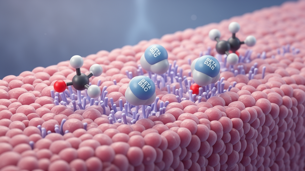
Nitric oxide and its generating enzymes occupy a pivotal junction between neuromodulation, oxidative stress, and immune activity. Under physiological conditions, neuronal and endothelial nitric oxide synthases generate baseline nitric oxide that acts as a retrograde messenger at glutamatergic synapses, activating soluble guanylate cyclase, cyclic guanosine monophosphate, and protein kinase G pathways to support long-term potentiation and memory. However, sustained induction of inducible nitric oxide synthase during inflammatory states yields excessive nitric oxide that rapidly reacts with superoxide anions to form peroxynitrite, a powerful oxidant that induces protein nitration, lipid peroxidation, DNA damage, and mitochondrial respiratory complex impairment. Patient findings regarding nitric oxide homeostasis remain complex and heterogeneous: Jiang and colleagues documented lower plasma total nitric oxide synthase, reduced inducible nitric oxide synthase, and higher malondialdehyde in 98 drug-naive first-episode patients, with total enzyme levels correlating positively with cognitive scores on the Repeatable Battery for the Assessment of Neuropsychological Status. Conversely, Lei and colleagues observed significantly elevated circulating nitric oxide metabolites alongside unaltered inducible enzyme mRNA in 58 patients, where nitric oxide levels trended negatively with cognitive performance without achieving statistical significance.
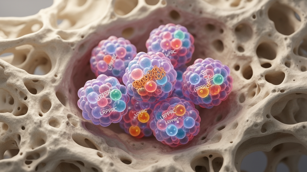
The authors analyzed how nitric oxide disruption could intersect with the canonical two-signal activation of the NLRP3 inflammasome, while clarifying that nitric oxide is not a universal inflammasome trigger. Signal 1 requires nuclear factor-kappa B-dependent priming to upregulate transcription of NLRP3, pro-interleukin-1 beta, and pro-interleukin-18, whereas Signal 2 involves stress-induced assembly triggered by potassium efflux, lysosomal destabilization, or mitochondrial reactive oxygen species and oxidized mitochondrial DNA. While peroxynitrite-induced mitochondrial respiratory chain damage can amplify mitochondrial reactive oxygen species to promote inflammasome assembly, nitric oxide can also exert inhibitory effects under alternative conditions by stabilizing mitochondria or directly mediating S-nitrosylation of NLRP3. Experimental cell studies demonstrate that cellular context dictates this response; mouse microglia possess full NLRP3 inflammasome assembly capacity, whereas patient-derived induced pluripotent stem cell astrocytes studied by Szabo and colleagues showed lower baseline NLRP3 and ASC expression but exaggerated caspase-1 activation and cytokine release upon direct stimulation, an inflammatory phenotype that was reversed by NLRP3 inhibition.
Crucially, the synthesis emphasized that inflammasome assembly, cytokine maturation and release, gasdermin D-mediated pore formation, and pyroptotic cell death represent related but distinct downstream events. Structural biology demonstrates that active caspase-1 cleaves full-length gasdermin D into an amino-terminal fragment that oligomerizes into plasma membrane pores, but viable macrophages can release interleukin-1 beta through these pores without undergoing immediate lysis. Consequently, detecting cytokine release or elevated transcripts does not prove pyroptotic cell destruction. In clinical studies, Lei and colleagues observed elevated peripheral NLRP3 mRNA in schizophrenia, which correlated negatively with verbal scores, and negative correlations between interleukin-18 mRNA and multiple cognitive domains, despite finding no significant group difference in interleukin-18 transcript levels between patients and controls. Furthermore, meta-analyses by Patlola and colleagues pooling 25 studies and 2,398 patients revealed that elevated peripheral interleukin-1 beta was negatively associated with attention, processing speed, visual learning, and working memory, though individual cognitive domain estimates relied on small patient subsets and peripheral assays rather than direct brain measurements.
To guide future experimental verification, the authors organized these multi-tiered findings into two alternative conceptual models: a serial regulation model and a parallel regulation model. The serial model hypothesizes that altered extracellular vesicle microRNAs dysregulate inflammatory networks, precipitating nitric oxide synthase imbalance and mitochondrial oxidative stress that lower the threshold for microglial NLRP3 activation, thereby impairing synaptic plasticity and contributing to cognitive deficits. In contrast, the parallel model posits that extracellular vesicle alterations, nitric oxide disturbances, and inflammasome priming arise concurrently from overarching metabolic and immune disruptions, with alternative pathways such as mTOR-dependent autophagy impairment independently modulating inflammasome activity. Resolving these hypotheses will require moving beyond static cross-sectional blood measurements toward single-cell transcriptomics, spatial mapping of cortical pathology, rigorous validation of vesicle cellular origins, and longitudinal studies evaluating whether source-resolved molecular panels can meaningfully stratify cognitive trajectories in schizophrenia beyond standardized neuropsychological assessments.
Subject of Research: A mechanistic hypothesis connecting extracellular vesicle microRNAs, nitric oxide signaling, and NLRP3 inflammasome activity to cognitive impairment in schizophrenia
Article Title: Extracellular vesicle miRNAs, NOS/NO homeostasis, and NLRP3 inflammasome signaling in schizophrenia-related cognitive impairment: a mechanistic review and testable hypothesis
Article References: Zhang, T., Tian, Y., Li, X., Zhang, H., & Liu, F. (2026). Extracellular vesicle miRNAs, NOS/NO homeostasis, and NLRP3 inflammasome signaling in schizophrenia-related cognitive impairment: a mechanistic review and testable hypothesis. Frontiers in Psychiatry, 17, Article 2013316. https://doi.org/10.3389/fpsyt.2026.2013316
Image Credits: AI Generated
DOI: 10.3389/fpsyt.2026.2013316
Keywords: schizophrenia, cognitive impairment, extracellular vesicles, microRNA, nitric oxide, nitric oxide synthase, NLRP3 inflammasome, neuroinflammation, microglia, synaptic pruning, pyroptosis, gasdermin D, interleukin-1beta, interleukin-18, mitochondrial dysfunction, oxidative stress
















 English (US) ·
 French (CA) ·