

 1 day ago
11
PROTECT YOUR DNA WITH QUANTUM TECHNOLOGY
Orgo-Life the new way to the future Advertising by AdpathwayA surveillance magnetic resonance imaging scan identified a previously unrecognized intracranial lesion in a fourteen-month-old boy ten months after surgical excision of a glabellar dermoid cyst, according to a pediatric case report and narrative literature review published in BMC Pediatrics. The initial clinical assessment had categorized the growth situated between the child’s eyebrows as a strictly extracranial anomaly. The subsequent intracranial finding demonstrates that subtle intracranial extensions of craniofacial cysts may escape primary radiological detection even when initial magnetic resonance imaging is performed prior to resection.
Epidermoid and dermoid cysts represent one of the most frequent benign skull and scalp tumors encountered in pediatric patients, accounting for 16 to 33 percent of all pediatric skull lesions. Both pathological entities develop from misfolded ectodermal inclusions during early embryonic development. While these benign congenital lesions commonly present as painless, indolent subcutaneous masses distributed around the orbit, they can also involve the surrounding periosteum and adjacent bone, occasionally progressing into deeper intracranial spaces.
Standard primary evaluation for pediatric subcutaneous lesions frequently begins with ultrasound imaging, wherein epidermoid and dermoid cysts characteristically present as well-circumscribed, avascular, round to oval, or lobulated masses containing echogenic keratin debris. Ultrasound examination, however, provides a reported sensitivity of only 50 percent for identifying intracranial spread. In contrast, magnetic resonance imaging demonstrates a reported sensitivity of 100 percent and a specificity of 95.7 percent, rendering advanced cross-sectional imaging necessary for lesions located along the cranial midline.
In the reported clinical case, the male infant presented with a glabellar mass present since birth that had gradually enlarged and repeatedly discharged white secretions. Physical examination identified a protruding lesion measuring three to four centimeters between the eyebrows, while initial laboratory investigations showed no systemic abnormalities. An initial ultrasound examination revealed an encapsulated, homogeneous, and poorly vascularized lesion within the soft tissues.
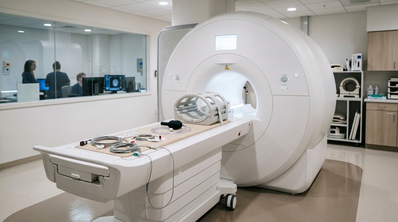
Institutional protocol prompted a non-contrast preoperative magnetic resonance imaging scan comprising T1-weighted, T2-weighted, and fluid-attenuated inversion recovery sequences alongside diffusion-weighted imaging and apparent diffusion coefficient maps. The scan revealed a fourteen-millimeter polylobulated extracranial mass displaying central T2 hyperintensity and peripheral T2 hypointensity. Initial neuroradiological evaluation identified no signs of intracranial communication or intracranial lesions.
An interdisciplinary tumor board reviewed the findings and scheduled complete neurosurgical resection of the extracranial mass two weeks later. During the initial surgical intervention, neurosurgeons observed that the mass adhered directly to the underlying cranial bone, where a central bony indentation contained connective tissue strands traversing into the cyst. Surgical teams coagulated and divided these fibrous strands, burred away the abnormal bone at the attachment site, and excised an overlying dermal sinus opening without exposing or opening the underlying dura mater.
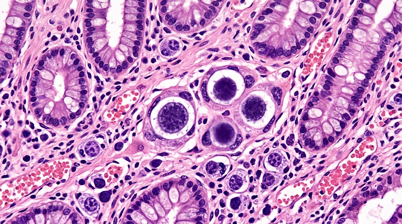
Histopathological analysis of the primary resected specimen confirmed a classic dermoid cyst lined by keratinizing stratified squamous epithelium accompanied by mature dermal appendages, including hair follicles and glandular components. Given the suspicious intraoperative bony findings, clinical teams recommended follow-up imaging at three months, though the patient’s family ultimately presented for surveillance magnetic resonance imaging ten months postoperatively.
The ten-month surveillance scan revealed an asymptomatic intracranial lesion situated in the right frontal parasagittal region characterized by restricted diffusion, with no recurrent tissue at the primary glabellar site. Retrospective re-examination of the initial baseline scans demonstrated that an extremely faint, subtle intracranial finding had been present preoperatively. This observation raised the clinical question of whether the intracranial component had gradually enlarged from a minute initial lesion or represented a postoperative recurrence.
Neurosurgical teams performed a secondary craniotomy in the right frontal region following contrast-enhanced magnetic resonance imaging, completely resecting the intracranial mass after opening the dura mater. No structural connection joined the lesion to the dura. Subsequent histopathological examination revealed epithelial fragments and abundant keratin lamellae lacking glandular structures, establishing a diagnosis of an epidermoid cyst.
A narrative review compiled by the authors identified 129 published pediatric cases featuring intracranial extension of dermoid or epidermoid cysts, with intracranial involvement noted in approximately 9 to 15 percent of reported childhood cases. Unchecked intracranial components carry risks of osteomyelitis, abscess formation, and meningitis. Follow-up imaging approximately four months after the second surgery showed no residual epidermoid tissue, and further surveillance examinations at 14 and 25 months post-resection confirmed complete clinical remission.
Understanding the distinct radiological features that differentiate epidermoid cysts from dermoid cysts on magnetic resonance imaging requires examining their internal microscopic composition. On magnetic resonance imaging, epidermoid cysts, which consist of layered keratin lamellae, appear isointense to cerebrospinal fluid across conventional acquisition sequences. In contrast, dermoid cysts appear characteristically hyperintense on T1-weighted images because their cyst walls contain additional skin appendages that continuously secrete an oily fluid directly into the cyst cavity. Because these two congenital cyst varieties are encountered across various medical disciplines, standardized recommendations for their diagnostic evaluation and therapeutic management remain necessary to establish definitive anatomical boundaries before surgical strategies are finalized.
Although epidermoid and dermoid cysts are benign, complete surgical excision is the therapy of choice and indicated for most pediatric patients. Unmanaged cysts can progressively enlarge or rupture, causing local inflammation, cosmetic issues, bone erosion, or neurological deficits when localized intracranially. Furthermore, cysts carry a rare possibility of malignant transformation into squamous cell carcinoma, which typically occurs during adulthood at an average age of 54 years and most often arises from intracranial lesions. Because recurrences following surgical care are very rare, clinical teams must individualize management based on lesion location and suspected extension, while maintaining surgical excision as the standard approach, especially for lesions situated along cranial midline structures.
Conducting neuroimaging in young pediatric patients involves institutional protocols and pharmacological precautions regarding sedation. Because intracranial lesions cannot be reliably excluded by ultrasound, the authors routinely perform magnetic resonance imaging in children presenting with skull lesions when intracranial involvement is possible, reviewing scans in their interdisciplinary tumor board. In this patient, the initial scan was performed under sedation with enterally administered chloral hydrate, regularly used in their institution. The authors emphasize that pediatric sedation requires an individualized assessment of clinical need, potential risks, and available alternatives, alongside appropriate monitoring and adherence to established safety standards. The report complied with the Declaration of Helsinki, with informed consent obtained from the patient’s legal guardian.
The selective administration of intravenous contrast agents introduces another essential diagnostic consideration when imaging pediatric cranial masses. Although contrast enhancement is not routinely required for dermoid and epidermoid cysts, contrast-enhanced sequences provide valuable diagnostic utility under specific clinical circumstances. In particular, the study authors note that intravenous contrast proves useful when atypical imaging features raise concern for alternative diagnoses or when an intracranial connection is suspected. Because craniofacial midline lesions carry the highest reported risk of intracranial extension, clinicians must exercise heightened vigilance when evaluating masses along the midline. Routine interdisciplinary discussion of such cases within a tumor board setting helps surgical teams accurately assess lesion extent, select tailored operative plans, and ensure appropriate treatment.
The divergent histopathological diagnoses rendered for the patient’s two surgical specimens provide valuable biological insight into the development and composition of congenital skull lesions. While the primary glabellar resection confirmed a dermoid cyst lined by keratinizing stratified squamous epithelium with mature dermal appendages including hair follicles and glandular structures, the secondary intracranial resection revealed numerous keratin lamellae and small fragments of epithelium without glandular components, establishing an epidermoid cyst. The authors suggest that this histological divergence may reflect the heterogeneous tissue architecture of a combined primary lesion. Because both entities originate from misfolded ectodermal inclusions during embryonic development, they may represent different manifestations of the same underlying tissue. The initial malformation may have contained both dermoid and epidermoid components that were unevenly distributed, causing each component to be represented differently within separate specimens. In addition, tissue fragmentation during surgery, biopsy sampling limitations, and the documented evolution of histological characteristics over time may have contributed to the discordant findings.
Anatomical patterns highlighted in the authors’ literature analysis also indicate that while intracranial extension occurs predominantly in craniofacial midline lesions, particularly frontonasal dermoid cysts, deep extension is definitely not restricted to midline structures. Published evidence documents intracranial extension in other anatomical skull locations, confirming that non-midline localization does not preclude intracranial involvement. However, the study authors caution that the unexpectedly high proportion of intracranial extension described across pediatric medical literature may partly reflect reporting bias, because uncomplicated dermoid cysts managed uneventfully are rarely reported. Immediate postoperative imaging provides vital baseline documentation following intracranial resection. In this patient, a postoperative magnetic resonance imaging scan performed one day after surgery revealed a resection cavity measuring up to 8 by 8 by 9 millimeters in the right frontal region, alongside an adjacent area in which a small residual lesion could not be excluded.
To prevent undetected disease progression, the authors recommend that follow-up magnetic resonance imaging 3 to 6 months after surgery should be considered whenever preoperative or intraoperative findings raise concern about possible deeper involvement or residual disease. The case illustrates that a subtle intracranial lesion may not be recognizable on initial image interpretation despite magnetic resonance imaging having been performed. Either progression of an extremely small initial lesion or intracranial recurrence remains the most likely explanation. Finally, the authors acknowledge that because this report represents a single case with a rare clinical course, conclusions must be confirmed by further evidence. Additionally, the literature mini-review was narrative rather than systematic, and no datasets were generated or analysed during the study.
Subject of Research: Intracranial extension of pediatric craniofacial dermoid and epidermoid cysts
Article Title: When a glabellar dermoid cyst is not the whole story: the significance of MRI in excluding intracranial segments
Article References: Wiedenhoefer, R., Feige, U., Shah, M., Gunst, L., Feuchtinger, T., & Puzik, A. (2026). When a glabellar dermoid cyst is not the whole story: the significance of MRI in excluding intracranial segments. BMC Pediatrics, 26(1), Article 1132. https://doi.org/10.1186/s12887-026-08016-6
Image Credits: AI Generated
DOI: 10.1186/s12887-026-08016-6
Keywords: dermoid cyst, epidermoid cyst, magnetic resonance imaging, pediatric neurosurgery, glabella, intracranial extension, case report, neuroradiology, glabellar, dermoid, cyst, whole
















 English (US) ·
 French (CA) ·