

 1 week ago
10
1 week ago
10
PROTECT YOUR DNA WITH QUANTUM TECHNOLOGY
Orgo-Life the new way to the future Advertising by AdpathwayThe part of the brain that handles language and reasoning and the part that keeps the body breathing appear to start from two different sets of embryonic cells, and the same two-part blueprint turns up in acorn worms, small burrowing animals that live on the seafloor. The finding comes from a Stanford Medicine-led study published September 18, 2026, in the journal Nature Neuroscience.
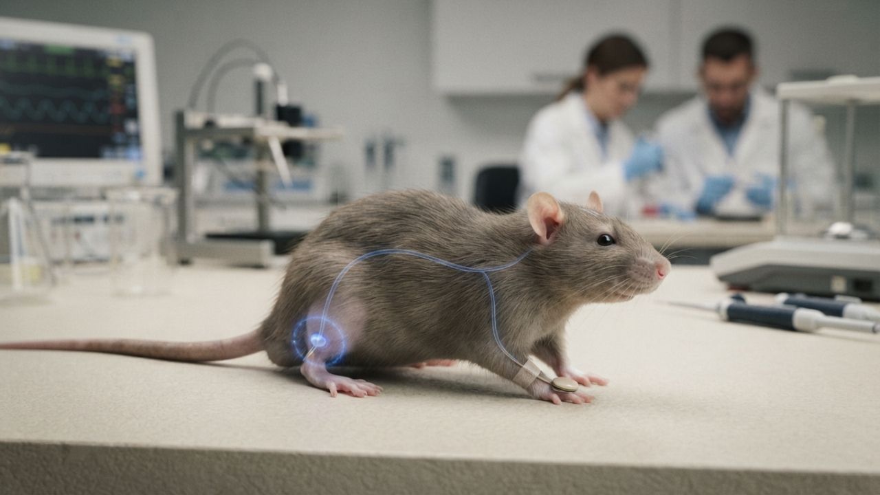
The practical payoff is in the lab. Using what they learned about early development, the researchers coaxed human stem cells into working hindbrain motor neurons for the first time. Those are the kinds of cells that fail in amyotrophic lateral sclerosis (ALS) and spinal muscular atrophy (SMA), diseases that can rob patients of the ability to swallow and breathe.
The study offers two things: a new window into how nervous systems evolved in animals from the seafloor to people, and a research tool that could help scientists study brain stem diseases.
Two Starting Cells, Two Brain Regions
The adult brain has three main regions. The forebrain handles higher-level thinking such as language and abstract reasoning. The hindbrain, which includes much of the brain stem, controls automatic functions such as breathing, sleep, heartbeat, and hunger, along with the muscles of the face, tongue, and throat.
For decades, the prevailing model held that a single type of early progenitor cell gave rise to the whole brain. According to Stanford Medicine's announcement of the two-part brain findings, co-first authors Rayyan Jokhai and Carolyn Dundes, graduate students in the lab of developmental biologist Kyle Loh, found otherwise by studying mouse embryos during gastrulation, the stage when the body plan first takes shape.
They identified two separate progenitor populations. Cells expressing a gene called Otx2 go on to form the forebrain and midbrain, while cells expressing Gbx2 form the hindbrain. The two groups never overlap, and their DNA is packaged in fundamentally different ways, which appears to lock each one onto its own developmental track.
That may explain decades of failed attempts to grow human hindbrain neurons. Jokhai said earlier efforts likely tried to turn forebrain and midbrain progenitors into hindbrain cells, which the study suggests is not possible.
A Blueprint Shared with Seafloor Animals
The team then looked across the animal kingdom. According to ScienceAlert, the same two-origin pattern appeared during early development in mouse, macaque, chicken, zebrafish, and acorn worm embryos, animals whose lineages scientists estimate diverged from a common ancestor around 550 million years ago. Acorn worms are soft-bodied marine animals that burrow into the seafloor and share a distant common ancestor with humans.
Loh said in the Stanford announcement that the research suggests "evolution took two existing neural systems and pushed them together spatially." He added that a single organ would probably be more efficient, but that animals rely on this ancient way of building the brain in two pieces.
Finding the pattern in acorn worms matters because these animals sit on a branch of the tree of life that separated from vertebrates very early. A shared blueprint suggests the two-part plan is old, predating fish, birds, and mammals. Stanford also noted that jellyfish, which diverged from the human lineage about 600 million to 700 million years ago, have two nervous systems at different ends of their bodies.
Headlines Versus Evidence
Stanford's own announcement described the brain as "two separate organs." That wording needs context. As ScienceAlert pointed out, the researchers do not mean the two neural systems were necessarily separate organs in some distant ancestor. The evolutionary story is an inference drawn from how modern embryos develop, and the brain still works as one connected organ.
The study's evidence comes from animal embryos and from human pluripotent stem cells grown in the lab, which followed the same two developmental paths when given the right signals. No human embryos were studied. ScienceAlert noted that the evidence cannot yet show whether the two parts actually evolved independently, a question left for future research.
Researchers from Caltech and the University of California, San Francisco contributed to the work, which was funded by the National Institutes of Health, the National Science Foundation, the California Institute for Regenerative Medicine, the Spinal Muscular Atrophy Foundation, and several private foundations, according to Stanford.
A New Tool for ALS and SMA Research
The most immediate impact is on disease research. Scientists cannot obtain brainstem tissue from living patients, and lab attempts to grow hindbrain neurons have long struggled. The Stanford team's lab-grown neurons produced action potentials, the electrical signals neurons use, and made proteins that mark the hindbrain segments controlling facial and swallowing muscles, according to an EurekAlert news release on the study.
SMA is a leading genetic cause of death in children under 1 year old, and ALS is often diagnosed between ages 40 and 70. In both, certain hindbrain neurons gradually stop working, which can lead to trouble swallowing, pneumonia from inhaled food or liquid, and eventually loss of the ability to breathe. Having these cells in a dish gives researchers a new model for studying what goes wrong.
This is early, basic research and does not offer a new treatment. Any therapy built on it would require years of further study and clinical trials. People living with ALS or SMA should continue to rely on their care teams for treatment decisions.
The researchers say their next steps are to trace the developmental origins of the spinal cord and to learn exactly how SMA and ALS damage hindbrain neurons. For evolutionary biologists, the open question is how and when the two systems came together, a puzzle that acorn worms and other early-branching animals may help answer.
What Readers Want to Know
What did the Stanford study find?
It found that the forebrain and midbrain develop from one group of embryonic progenitor cells and the hindbrain from a separate group, and that the two groups never overlap.
What are acorn worms?
Acorn worms are small, soft-bodied marine animals that burrow into the seafloor. They share a distant common ancestor with vertebrates, including humans.
Is the human brain really two separate organs?
Not in the everyday sense. The brain works as one connected organ, but its front and back regions arise from different starting cells. The evolutionary history is an inference.
Why does this matter for ALS and SMA?
The team grew human hindbrain motor neurons in a dish for the first time, giving researchers a new way to study cells damaged in these diseases.
Does this lead to a new treatment?
No. It is early laboratory research. Any therapy based on it would need years of additional study and clinical trials.
Where was the study published?
It was published on September 18, 2026, in Nature Neuroscience, led by Stanford Medicine researchers with collaborators at Caltech and UCSF.
© 2026 NatureWorldNews.com All rights reserved. Do not reproduce without permission.

















 English (US) ·
  French (CA) ·