

PROTECT YOUR DNA WITH QUANTUM TECHNOLOGY
Orgo-Life the new way to the future Advertising by Adpathway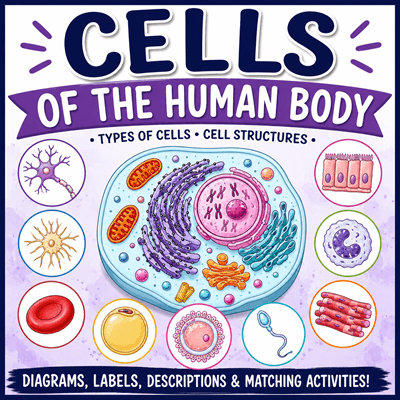
Every living organism is made of cells, but not all cells look—or function—the same. One of the biggest challenges for students in biology and anatomy is understanding how a single basic cell plan can be modified into hundreds of specialized cell types. This Cells of the Human Body Coloring and Matching Activity helps students make those connections through coloring, observation, and review.
Rather than memorizing vocabulary from a textbook, students actively explore the structures that make up a typical animal cell while comparing it to specialized cells found throughout the human body.
Why Teach Cell Diversity?
Students are often introduced to the “typical” animal cell early in biology, but they may not realize that muscle cells, neurons, blood cells, and bone cells all share many of the same organelles. Each specialized cell has adapted its shape and structure to perform a specific job, while still carrying out the basic functions of life.
This activity emphasizes that relationship by placing a detailed animal cell in the center of the worksheet and surrounding it with examples of specialized human cells. Students can immediately compare the unique shapes of each cell while recognizing that they all originated from the same basic cellular blueprint.
A Closer Look at Cell Organelles
The centerpiece of the worksheet is a detailed animal cell that students color while identifying its major organelles. As they work through the diagram, they review the functions of important cellular structures, including:
- Plasma membrane – forms the outer boundary of the cell
- Cytoplasm – fluid that fills the cell
- Nucleus – the cell’s control center
- Nucleolus – inside the nucleus; produces ribosomes
- Rough endoplasmic reticulum – helps produce, fold, and transport proteins
- Smooth endoplasmic reticulum – produces lipids, breaks down harmful substances
- Ribosomes – build proteins
- Golgi apparatus – modifies, sorts, and packages proteins
- Mitochondria – the cell’s “powerhouses;” they produce ATP
- Lysosomes – contain digestive enzymes that break down substances
- Centrioles – play an important role during cell division
Coloring encourages students to slow down and carefully examine each structure rather than simply labeling a diagram. Many teachers find that students remember organelles more effectively after assigning each one a different color.
Meet the Specialized Cells
Surrounding the central cell are illustrations of several specialized human cells that students encounter throughout a biology or anatomy course.
The activity includes:
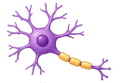
- Neuron (nerve cell)
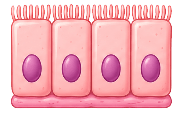
- Epithelial cell
- Osteocyte (bone cell)
- White blood cell
- Skeletal muscle cell
- Red blood cell
- Fat cell (adipocyte)
- Egg cell (ovum)
- Sperm cell
Reinforce Learning with Matching Activities
After completing the coloring page, students can test their understanding using two matching exercises.
The first reviews cell organelles, requiring students to match each structure with its function. This helps students distinguish between similar organelles such as the rough and smooth endoplasmic reticulum or the Golgi apparatus and lysoso
Related Activities
Microscope Introduction with E Slides – learn the basics of the microscope
Exploring Different Types of Cells – students view different types of cells under the microscope
Lab: Comparing Human and Frog Blood Cells – View both types of cells under the microscope, noting the key difference is that frog blood cells have a nucleus
Slides and Student Notes on Cell Biology – Anatomy focused slides on the cell unit, with a case story about primary ciliary dyskinesia
Giant Microbe Cell Plushes – this set has most of the body cells, add them to your lecture for a visual reference!

















 English (US) ·
  French (CA) ·