

PROTECT YOUR DNA WITH QUANTUM TECHNOLOGY
Orgo-Life the new way to the future Advertising by AdpathwayArticle navigation

RESEARCH ARTICLE| 02 July 2026
Niclas U. Lundsgaard
,
Niclas U. Lundsgaard *
Conceptualization, Data curation, Formal analysis, Investigation, Methodology, Project administration, Visualization, Writing – original draft, Writing – review & editing
School of the Environment,
The University of Queensland
,
Brisbane, QLD 4072
,
Australia
Search for other works by this author on:
Craig E. Franklin
,
Craig E. Franklin
Conceptualization, Funding acquisition, Project administration, Resources, Supervision, Writing – review & editing
School of the Environment,
The University of Queensland
,
Brisbane, QLD 4072
,
Australia
Search for other works by this author on:
Rebecca L. Cramp
Rebecca L. Cramp
Conceptualization, Funding acquisition, Methodology, Resources, Supervision, Visualization, Writing – review & editing
School of the Environment,
The University of Queensland
,
Brisbane, QLD 4072
,
Australia
Search for other works by this author on:
*
Present address: Water Planning Ecology, Queensland Department of the Environment, Tourism, Science and Innovation, Dutton Park, QLD 4102, Australia.
Competing interests
C.E.F. is the Editor in Chief of the Journal of Experimental Biology. C.E.F. was not involved in the editorial assessment of this submission. The authors declare no other competing interests.
Received: 21 Jan 2026
Accepted: 15 May 2026
Online ISSN: 1477-9145
Print ISSN: 0022-0949
Funding
Funding Group:
Award Group:
- Funder(s):
Australian Research Council
- Award Id(s):
DP190102152
- Funder(s):
Funding Group:
Award Group:
- Funder(s):
Australian Government
- Funder(s):
Funding Group:
Award Group:
- Funder(s):
The University of Queensland
- Funder(s):
© 2026. Published by The Company of Biologists
2026
This is an Open Access article distributed under the terms of the Creative Commons Attribution License (https://creativecommons.org/licenses/by/4.0), which permits unrestricted use, distribution and reproduction in any medium provided that the original work is properly attributed.
J Exp Biol (2026) 229 (13): jeb252227.
ABSTRACT
Accumulation of unrepaired DNA lesions is a key mechanistic driver of mortality in organisms exposed to elevated ultraviolet-B radiation (UVBR). Recent research has found that high irradiance UVBR causes disproportionate mortality in larvae of a UV-sensitive amphibian (Limnodynastes peronii) compared with an equivalent dose of low irradiance UVBR. Here, we aimed to determine whether this irradiance-dependent effect is also reflected in the DNA damage profile of L. peronii larvae. Under laboratory conditions, we exposed larvae to one of two different UVBR irradiances over 2 days, keeping the daily dose equal by adjusting exposure duration. Larvae were sampled at set time points throughout experimental exposures for quantification of cyclobutane pyrimidine dimer (CPD) concentration. Larvae were highly susceptible to UVBR-induced CPD formation and had low rates of DNA repair, resulting in a carryover of DNA damage into the following day. Consequently, CPD concentrations accumulated to a higher level during the second day of exposure, an effect that was exacerbated by high irradiance exposure. Larvae exposed to high irradiance UVBR accumulated CPD lesions almost 3 times faster than larvae exposed at half the irradiance, causing a 47% increase in CPD concentration across sampling time points. These results demonstrate the capacity for high irradiance exposure to exacerbate UVBR-induced genomic instability, which may explain the irradiance-dependent mortality observed in this species. Given the conserved nature of DNA repair mechanisms across taxa, these findings have implications for other aquatic organisms with a larval life stage, including fish and coral, that are facing increasing UVBR levels owing to climate change.
INTRODUCTION
Solar ultraviolet radiation (UVR) is a ubiquitous environmental stressor that impacts essential biomolecules (Fraikin et al., 2024), organisms (Downie et al., 2023) and ecosystems (Williamson et al., 2019). Among the three UVR wavelength bands (UVAR: 315–400 nm, UVBR: 280–315 nm and UVCR: 100–280 nm), UVBR exerts the greatest influence on the biosphere (de Gruijl, 2002; Fraikin et al., 2024). UVBR photons are directly absorbed by DNA, causing lesions in the form of cyclobutane pyrimidine dimers (CPDs) and pyrimidine–pyrimidone photoproducts (6-4PPs; Sinha and Häder, 2002; Fraikin et al., 2024). These lesions disrupt transcription and replication of DNA, which can trigger cell death (Campisi and d'Adda di Fagagna, 2007; Batista et al., 2009). If photo-damaged cells are not eliminated, then mutations, tumours and malformations can result (Batista et al., 2009; Cadet and Douki, 2018; Fraikin et al., 2024).
UVBR-induced genomic instability can have a variety of downstream consequences on performance, behaviour and survival across taxa (Bancroft et al., 2007; Llabrés et al., 2013; Peng et al., 2017; Downie et al., 2023). Amphibians are particularly vulnerable because they lack UV-protective feathers and fur. Additionally, amphibians are typically exposed to UVBR during embryonic and larval stages (Bancroft et al., 2008a; Downie et al., 2023). This is a time of rapid cell division and significant developmental milestones, making them particularly sensitive to UVBR-induced DNA damage and methylation changes (Londero et al., 2019; de Oliveira et al., 2020). Like most taxa, amphibians possess DNA repair mechanisms including nucleotide excision repair (NER) and photoreactivation to alleviate DNA damage (Sinha and Häder, 2002; Rastogi et al., 2010). Photoreactivation specifically targets and reverses UV-induced pyrimidine dimers with DNA photolyase enzymes that use light energy in the UVAR and visible spectra (300–500 nm) (Sancar, 2008; Rastogi et al., 2010). Unsurprisingly, amphibian species with lower DNA repair efficiencies generally suffer higher UV-induced mortality rates (UV sensitivity hypothesis; Blaustein et al., 1994; Hays et al., 1996).
Elevated UVBR has been implicated in amphibian population declines since the onset of stratospheric ozone depletion in the 1980s (Kiesecker and Blaustein, 1995; Lips, 1998; Kiesecker et al., 2001; Alton and Franklin, 2017). However, the magnitude of this cause–effect relationship remains a mystery, due in part to a scarcity of dose–response data needed for informing population modelling (Diamond et al., 2002; Beebee and Griffiths, 2005; Olker et al., 2013; Hird et al., 2025b). Elucidating how the key parameters of UVBR exposure influence physiological responses of amphibians is crucial, including the effects of dose, irradiance and diurnal pattern of exposure. This information can inform experimental design, ecological interpretation and predictive models of UV exposure in natural systems (Lundsgaard et al., 2021). Laws and principles developed in photochemistry provide a useful framework for addressing this knowledge gap.
The Bunsen–Roscoe Law of Reciprocity (BRL) posits that photochemical effects are proportional to the dose of radiation, regardless of the combination of irradiance and exposure duration [effect∝dose (kJ m−2)=irradiance (μW cm−2)×exposure duration (s)] (Bunsen and Roscoe, 1855). For example, in vitro exposure of purified DNA to UVBR generates lesions in a dose-dependent and irradiance-independent manner (reflecting a linear relationship with energy input; Schindl et al., 2001; Pandelova et al., 2006). Studies investigating UVBR administration as a form of agricultural pest control have often found the BRL to hold true for photobiological responses in a range of invertebrate pest species (Sakai et al., 2012; Murata and Osakabe, 2013; Parajuli et al., 2023; Holford et al., 2024). However, the BRL has limited utility in predicting other biological outcomes in vivo, such as UVBR-induced tumorigenesis and skin cancer progression (Kelfkens et al., 1991; Schindl et al., 2001; Lan et al., 2016), and UVBR-induced mortality in larval amphibians (Lundsgaard et al., 2021, 2022, 2025).
In contrast to the BRL, we previously found that a multi-day exposure of striped marsh frog larvae (Limnodynastes peronii) to high irradiance UVBR (38.6 μW cm−2) was 3 times more lethal than an equivalent dose administered at lower irradiance (9.3 μW cm−2; Lundsgaard et al., 2025). One hypothesis for this irradiance-dependent sensitivity is that in vivo responses including DNA repair are only able to protect against the accumulation of DNA damage up until a certain irradiance toxicity threshold. Beyond this point, DNA lesions are generated at a rate that exceeds repair capacity, resulting in an accumulation of DNA damage in the genome and subsequent mortality (Pandelova et al., 2006). No studies to our knowledge have examined the DNA damage profile underlying such irradiance-dependent effects by explicitly controlling for dose.
The lethal effects of high irradiance UVBR can often take multiple consecutive days of exposure to manifest, suggesting a gradual, multi-day degradation of genomic integrity (Tietge et al., 2001; Pandelova et al., 2006; Lundsgaard et al., 2025). Whether the accumulation of DNA damage over time is the result of a daily carry-over of unrepaired DNA or a reduction in DNA repair rates over time is yet to be determined (Pandelova et al., 2006). Laboratory studies have indeed observed remnant DNA damage persisting in amphibian larvae the day after a UVBR exposure event (Schuch et al., 2015b), including in L. peronii (Morison et al., 2020; Hird et al., 2022). However, these studies did not incorporate naturalistic diurnal cycles, an important consideration given that photoreactivation is inactive in the absence of light at night (Rastogi et al., 2010). Evidently, advancing knowledge of the physiological consequences of different UVBR exposure regimes in amphibians is a necessary step in improving experimental designs, ecological interpretation of field measurements and modelling of UVBR-induced amphibian population declines.
Our aim was to characterise the DNA damage profile in L. peronii larvae induced by different UVBR exposure regimes administered over consecutive days under an ecologically appropriate diurnal light cycle (12 h light:12 h dark photoperiod regime). We exposed L. peronii larvae to different UVBR irradiances and measured the time course of CPD concentrations by sampling animals at set time points throughout a 2 day exposure period. Based on the irradiance-dependent mortality observed in our previous study (Lundsgaard et al., 2025), we hypothesised that the BRL would not apply and that high irradiance UVBR would cause greater concentrations of DNA damage in exposed larvae compared with an equivalent dose of low irradiance UVBR. We also predicted that DNA damage would carry over into the following day, and that larvae receiving a second day of UVBR exposure would consequently have higher concentrations of DNA damage compared with larvae exposed for only 1 day.
MATERIALS AND METHODS
Animal ethics approval
This research was approved by The University of Queensland Animal Ethics Committee (approval no. SBS/089/19) and animal collection permission was granted by the Queensland Department of Environment and Science (permit no. WISP17421516).
Study species
Limnodynastes peronii (Duméril and Bibron 1841) is a common species listed as ‘Least Concern’ by the IUCN, occurring along the eastern coast of Australia from northern Queensland down to the island of Tasmania (16–42°S; Hero et al., 2004; Anstis, 2017). This species is the primary model organism in Australia for research on the physiological effects of UVBR on amphibians and is characterised by a relatively low tolerance to UVBR exposure (van Uitregt et al., 2007; Alton et al., 2010; Bernal et al., 2011; Lundsgaard et al., 2020, 2025; Morison et al., 2020; Hird et al., 2022). This species breeds at most times of the year in shallow (often ephemeral) waterbodies that form after rain, including exposed, flooded grasslands that lack a tree canopy (Anstis, 2017). Egg clutches are laid as floating foam masses amongst vegetation and newly hatched larvae remain at the water surface for up to 2 days prior to their free-swimming feeding stage (N.U.L., personal observation). Larvae of this species are primarily diurnal and bottom dwelling, but they swim throughout the water column when feeding (Anstis, 2017). It is not known whether larvae of this species seek shady environments to avoid UVBR or prefer warmer, sunlit waters to thermoregulate at the cost of harmful UVBR exposure (Bancroft et al., 2008b). This species has been recorded in waterbodies where UVBR levels reach up to 500 μW cm−2 at the water surface (van Uitregt et al., 2007).
Animal collection and husbandry
A portion of each of five L. peronii egg masses was collected, the morning after laying, from a flooded creek bank in Brisbane, Australia (−27.58054S, 153.07616E). The clutches were transported immediately to The University of Queensland, where they were individually housed in 2 l plastic containers in a mixture of carbon-filtered Brisbane city tap water and water from the natal water body. Eggs were reared at 25°C under a 12 h light:12 h dark photoperiod regime using non-UVBR fluorescent ceiling lights. Upon hatching, larvae were evenly distributed amongst 16, 2 l plastic containers. Larvae were fed thawed spinach ad libitum throughout their development, and water quality was maintained with regular 80% water changes, adjusted to pH 6 with 10% hydrochloric acid. Fraction (Continuum Aquatics Fraction, Fort Payne, AL, USA) was added with each water change as per the manufacturer's instructions to detoxify nitrogenous waste.
After 3 weeks of development, the temperature was adjusted to 22°C for the remainder of the experiment. Larvae were given 9 days to adjust to the new temperature, at which point they were randomly distributed amongst 45 transparent plastic treatment containers (1 l; 15×10×7 cm) with a water depth of 5 cm. In total, there were nine containers per treatment with eight tadpoles per container (n=72 per treatment). Only a subset of these animals were sampled for the assay reported on in this study.
Experimental design
UVBR exposure
Larvae were given 5 days to adjust to experimental housing conditions prior to treatment exposure. Initial exposure to UVBR can elicit a ‘UV-hardening’ response that influences the tolerance of amphibian larvae to subsequent UVBR exposure (Calfee et al., 2006; Bancroft et al., 2008a). As such, experimental larvae (now 5 weeks old, Gosner stage 25–26; Gosner, 1960) were exposed to a low dose (0.293 kJ m−2) of ‘pre-treatment’ UVBR intended to standardise the UV-induced physiological state of larvae across treatments without causing major sub-lethal effects (Lundsgaard et al., 2025). This initial low dose of UVBR was administered at a low irradiance in the 2 days preceding treatment exposure (8.1±0.9 μW cm−2 for 30 min per day, from 13:00 h to 13:30 h). A separate group of animals (n=72) serving as a negative control did not receive this initial UVBR exposure and were sampled immediately prior to experimental treatments, euthanised in buffered tricaine-S (MS-222; Aqua-Life, Nanaimo, BC, Canada; 0.5 g l−1) and snap-frozen at −80°C.
After the 2 day pre-treatment exposure, larvae were exposed to one of four UVBR treatments in a fully factorial design experiment. There were two levels of UVBR irradiance (low 40.1 μW cm−2 and high 80.3 μW cm−2) and two levels of UVBR dose [low ∼2.9 kJ m−2 (1 day of exposure); and high ∼5.8 kJ m−2 (2 days of exposure)]. These levels were selected based on previous research demonstrating substantial but non-lethal effects of similar acute exposure regimes on this species (Hird et al., 2022, 2023, 2024; Lundsgaard et al., 2025). The UVBR levels are well below the ambient levels that occur in the region where the animals were collected (500 μW cm−2 at water level during midday; van Uitregt et al., 2007) to account for the likely attenuative effects of environmental factors such as vegetation cover and dissolved organic matter (Olker et al., 2013; Alton and Franklin, 2017; Santos et al., 2023). Daily UVBR dose was kept equal across irradiance treatments by adjusting the exposure period, with animals in the low irradiance treatment exposed for 2 h per day (12:00–14:00 h) and animals in the high irradiance treatment exposed for 1 h per day (12:15–13:15 h; Table 1). The start time of exposure was staggered by 15 min between treatments to allow time for sampling (Fig. 1).
Fig. 1.

A time line of the experimental treatments and sampling points in this study. The yellow bars represent the UVBR exposure periods of Limnodynastes peronii larvae for each irradiance treatment of the 2 day experiment. Note that all animals sampled on day 2 were exposed to the day 1 UVBR exposure regime, including the day 2 pre-exposure samples. There were six sampling time points in total: pre-exposure (solid lines), immediately after exposure (dotted lines) and 3 h after exposure (dashed lines) across both day 1 (blue lines) and day 2 (red lines) of experimentation. UVBR dose (approximately 2.9 kJ m−2 per day) remained constant across the low and high irradiance treatments. The grey bar represents the 12 h dark period in the diurnal cycle. The 2 day pre-treatment UVBR exposure is excluded from this diagram for simplicity. Created with BioRender by Lundsgaard, N., 2026. https://BioRender.com/xn1iifq. This figure was sublicensed under CC-BY 4.0 terms.
Fig. 1.
A time line of the experimental treatments and sampling points in this study. The yellow bars represent the UVBR exposure periods of Limnodynastes peronii larvae for each irradiance treatment of the 2 day experiment. Note that all animals sampled on day 2 were exposed to the day 1 UVBR exposure regime, including the day 2 pre-exposure samples. There were six sampling time points in total: pre-exposure (solid lines), immediately after exposure (dotted lines) and 3 h after exposure (dashed lines) across both day 1 (blue lines) and day 2 (red lines) of experimentation. UVBR dose (approximately 2.9 kJ m−2 per day) remained constant across the low and high irradiance treatments. The grey bar represents the 12 h dark period in the diurnal cycle. The 2 day pre-treatment UVBR exposure is excluded from this diagram for simplicity. Created with BioRender by Lundsgaard, N., 2026. https://BioRender.com/xn1iifq. This figure was sublicensed under CC-BY 4.0 terms.
Close modal
Table 1.
Ultraviolet-B (UVBR) and ultraviolet-A (UVAR) irradiances and dose estimates for each treatment
| Low (2 h day−1) | 40.1±1.9 | 2.888±0.137 | 5.776±0.274 | 216.8±21.0 | 15.612±1.513 | 31.224±3.026 |
| High (1 h day−1) | 80.3±3.2 | 2.890±0.115 | 5.780±0.230 | 504.3±36.2 | 18.154±1.304 | 36.308±2.608 |
Dose estimates were calculated based on the UVBR irradiance measurements and exposure durations, where low dose represents 1 day of exposure and high dose represents 2 days of exposure. Values are presented as means±s.e.m.
UVBR conditions were generated with 1.2 m, 40 W full spectrum fluorescent light tubes (Repti-Glo 10.0, Exo Terra, Montreal, QC, Canada). The low irradiance treatment shelf had six tubes mounted 27 cm above the water surface of treatment containers, whilst the high irradiance treatment had eight tubes mounted 12 cm above the water surface. UVBR and UVAR levels were measured at the water surface of each container using a photometer (IL1400BL, International Light Inc., Newburyport, MA, USA) to ensure consistent levels across experimental shelves (Table 1). In the field, UVBR and UVAR levels are somewhat correlated and follow similar diurnal patterns (Santos et al., 2023), which is important given the role of UVAR in photorepair of UVBR-induced DNA lesions (Sancar, 2008). Thus, the ratio of UVBR:UVAR was kept as similar as possible between experimental treatments, allowing for photorepair to occur in a proportionate manner across treatments. Additional non-UVR room lighting provided low levels of photoreactive light during 12 h daytime periods.
Temperature
Water temperature was measured during the experiment using waterproof iButton temperature data loggers and was 20.5±0.5°C in the low irradiance treatment and 21±0.5°C in the high irradiance treatment. Temperature changes during UVBR exposure periods were a 2°C increase for the low irradiance treatment, while the high irradiance treatment was limited to a 1°C increase by inserting trays of ice placed on ‘buffer shelves’ immediately underneath the treatment shelving during exposure periods. As such, irradiance treatments typically differed by 1°C or less.
Sampling regime
The experimental treatment period spanned 2 days, with three sampling time points each day (Fig. 1). These time points were immediately pre-UVBR exposure, immediately post-UVBR exposure and 3 h post-exposure (Fig. 1). Six tadpoles per treatment were sampled (from different replicate containers) at each time point for assessment of DNA damage. Additionally, two reference animals that did not receive the initial UVBR pre-treatment served as a negative control and were sampled prior to experimentation. Sampled individuals were immediately euthanised in MS-222 under red light (to prevent DNA photo-repair), then blotted dry and snap-frozen at −80°C.
Measuring CPD concentration
We restricted our investigation to CPDs, which are the most common and cytotoxic UV-induced DNA lesion, constituting 75–90% of DNA photoproducts (Sinha and Häder, 2002; Sancar, 2008; Fraikin et al., 2024). Genomic DNA was extracted and purified from the tissue of 74 animals (n=6 per time point, plus two no-UVBR reference animals), using a PureLink Genomic DNA Mini Kit (Invitrogen, Carlsbad, CA, USA), in accordance with the manufacturer's protocol for mammalian tissue. Seventeen larvae (across all treatments) were too large to process in their entirety, so these were eviscerated prior to use. DNA concentrations were quantified using a Qubit fluorometer (Invitrogen, cat. no. Q32857) and a Qubit 1× dsDNA HS Assay Kit (Invitrogen, cat. no. Q33230), in accordance with the manufacturer’s protocol. DNA samples were aliquoted and stored at −20°C until use.
CPD concentrations were quantified fluorometrically using an anti-CPD ELISA assay following the primary antibody manufacturer's protocol (Mori et al., 1991; NM-ND-D001, clone TDM-2, Cosmo Bio Co., Ltd, Tokyo, Japan) and following Hird et al. (2023). All reactions were prepared in triplicate across three clear flat-bottom 96-well plates (Thermo Fisher Scientific MaxiSorp Nunc-Immuno Plate, Roskilde, Denmark, cat. no. 442404) pre-coated in 0.003% protamine sulfate; 50 μl of each DNA sample (0.4 ng μl−1) and UVC-irradiated standards (0, 2.5, 5, 7.5, 10 J m−2; Cosmo Bio Co., Ltd, cat. no. NM-MA-R010; concentration 0.4 ng μl−1) per reaction were added to each plate. A PBS sample was added as a blank to detect background absorbance on each plate. Absorbance was measured at 450 nm (Beckman Coulter DTX880 multimode detector, Chaska, MN, USA). Data were acquired using the SoftMax® Pro program (version 7.1.0, Molecular Devices LLC, San Jose, CA, USA). All absorbance values were corrected for background absorbance. The interpolation curve was developed in Microsoft Excel by plotting a second-order polynomial trend line through the absorbance values for the standards (x-axis) against their known CPD concentration (y-axis). The equation generated from the trend line was used to determine CPD concentration (UVC-dose equivalent/20 ng DNA), of each sample based on the absorbance value.
Statistical analysis
Three statistical analyses of CPD concentration were conducted in the R statistical environment (version 4.1.1, ‘Kick Things’, http://www.R-project.org/). Regression diagnostics were performed using the performance package (Lüdecke et al., 2021). Models assumed a Gaussian error structure and satisfied assumptions of tests. Stepwise model selection with Akaike information criteria (AIC) was used to identify the best models. AIC showed there were no detectable container effects, so this random effect variable was excluded from all models. Significant effects between treatments were further elucidated with post hoc pairwise comparisons using the estimated marginal means package (emmeans), applying Tukey's HSD method to adjust for multiple comparisons (function emmeans, https://CRAN.R-project.org/package=emmeans).
Rather than reporting ‘statistical significance’ in the traditional sense, P-values were interpreted using a ‘language of evidence’ approach that better reflects the P-value as a continuous probabilistic measure of statistical evidence. That is, no evidence when P>0.1, weak evidence when 0.1≤P>0.05, moderate evidence when 0.05≤P>0.01, strong evidence when 0.01≤P>0.001 and very strong evidence when P≤0.001 (Muff et al., 2022). This method avoids problems associated with binary significance testing based on an arbitrary P-value threshold (Muff et al., 2022). Data are presented as means±s.e.m.
CPD concentrations in L. peronii larvae were predicted by three sets of categorical variables: sampling time point (3 levels), day/dose (2 levels) and irradiance (2 levels). Body mass was also included as a continuous covariate, thus requiring ANCOVA analysis. Given that day 1 pre-exposure samples were not exposed to UVBR, inclusion in a model with post-exposure treatments could skew treatment comparisons. As such, the pre-exposure and post-exposure sampling events were best represented as two distinct datasets analysed with separate models (analyses 2 and 3). Additionally, the effect of the 2 day UVBR pre-treatment exposure on baseline CPD concentrations was analysed (analysis 1).
Analysis 1 – pre-treatment CPD concentration
Negative control animals reared in the complete absence of UVBR had no detectable CPDs. Thus, the presence of CPDs in day 1 pre-exposure samples was analysed using a one-sample, one-sided t-test. The null hypothesis was that the population mean of CPD concentration was equal to zero and the alternative hypothesis was that the population mean was greater than zero.
Analysis 2 – post-exposure time points
A three-way Type III ANCOVA was used to model the influence of UVBR irradiance, day/dose, time point and mass on CPD concentration for the post-exposure sampling events (end of exposure versus 3 h post-exposure) (lme4 package, function lmer;Bates et al., 2015). This analysis compared CPD concentration in response to irradiance at various sampling time points over consecutive days of UVBR exposure, thus addressing hypotheses related to DNA repair rates and the irradiance-dependent accumulation of DNA damage.
Analysis 3 – pre-exposure time points
A two-way Type III ANCOVA was used to model the influence of UVBR irradiance, day (1 versus 2) and mass on CPD concentration between the two pre-exposure sampling events (lme4 package, function lmer; Bates et al., 2015). Day 1 pre-exposure samples were not exposed to UVBR, whilst day 2 pre-exposure animals were exposed to the various UVBR treatments on day 1 and then sampled the following day, prior to day 2 exposures. Thus, this analysis explicitly tested whether UVBR-induced DNA damage can carry over into the following day under a diurnal light cycle.
Predicted values were extracted from both ANCOVA models using the effects package for assessment of effect sizes (Fox and Weisberg, 2019).
RESULTS
Analysis 1 – pre-treatment CPD concentration
Control animals reared in the complete absence of UVBR had no detectable CPDs. There was, however, very strong evidence of residual CPDs (approximately 0.18 units) in day 1 pre-exposure animals, induced by the initial low irradiance UVBR pre-treatment exposure (t11>5.749, P<0.001; Fig. 2A).
Fig. 2.

Effect of UVBR exposure regime on DNA damage in L.peroniilarvae. DNA damage was quantified as the concentration of cyclobutane pyrimidine dimers (CPDs) in whole-body samples (calculated as UVC-dose equivalent/20 ng DNA). (A) The full dataset, with UVBR irradiance treatment on the x-axis graphed against three sampling time points [pre-exposure, immediately after exposure (‘end of exposure’) and 3 h post-exposure] over a 2 day exposure period (day 1, blue; day 2, red; n=6 per time point per treatment). (B–D) Post-exposure data for UVBR irradiance (B), exposure day (dose; C) and sampling time point (D), pooled across all other factors to illustrate main effects (in the absence of interactions between factors). Data points show individual animals (n=24 per group). Error bars indicate means±s.e.m., and asterisks denote statistically significant differences (Type III ANCOVA, P≤0.01).
Fig. 2.
Effect of UVBR exposure regime on DNA damage in L.peroniilarvae. DNA damage was quantified as the concentration of cyclobutane pyrimidine dimers (CPDs) in whole-body samples (calculated as UVC-dose equivalent/20 ng DNA). (A) The full dataset, with UVBR irradiance treatment on the x-axis graphed against three sampling time points [pre-exposure, immediately after exposure (‘end of exposure’) and 3 h post-exposure] over a 2 day exposure period (day 1, blue; day 2, red; n=6 per time point per treatment). (B–D) Post-exposure data for UVBR irradiance (B), exposure day (dose; C) and sampling time point (D), pooled across all other factors to illustrate main effects (in the absence of interactions between factors). Data points show individual animals (n=24 per group). Error bars indicate means±s.e.m., and asterisks denote statistically significant differences (Type III ANCOVA, P≤0.01).
Close modal
Analysis 2 – post-exposure time points
There was strong statistical evidence for independent effects of irradiance, dose and sampling time point on the CPD concentration of L. peronii larvae sampled at the post-exposure time points (irradiance: F1,43=32.995, P<0.001; dose: F1,43=7.196, P=0.010; sampling time point: F1,43=27.159, P<0.001; Fig. 2A). However, there was no evidence of interactive effects between any factors. There was strong evidence for an inverse relationship between body mass and CPD concentration (F1,43=7.198, P=0.010; Fig. 3). The model prediction estimated that an animal with the smallest mass in the sample population would have approximately 50% greater CPD concentration than the largest animal in the sample population.
Fig. 3.

Correlation between CPD concentration and mass of L. peronii larvae exposed to UVBR treatments. Data points represent individual animals, sampled at the end of exposure and 3 h post-exposure time points (n=48). The trend line represents the prediction from the model ([CPD]=3.75Irradiance−3.33Time+1.70Day−0.09Mass+10.63+ε) with all variables other than mass standardised. Dashed lines represent the standard error of the model prediction.
Fig. 3.
Correlation between CPD concentration and mass of L. peronii larvae exposed to UVBR treatments. Data points represent individual animals, sampled at the end of exposure and 3 h post-exposure time points (n=48). The trend line represents the prediction from the model ([CPD]=3.75Irradiance−3.33Time+1.70Day−0.09Mass+10.63+ε) with all variables other than mass standardised. Dashed lines represent the standard error of the model prediction.
Close modal
There was very strong evidence for an irradiance-dependent effect on CPD concentration, with CPDs accumulating 2.9 times faster during exposure to high irradiance UVBR compared with low irradiance UVBR (assuming a linear rate of accumulation). Consequently, the CPD concentration of L. peronii larvae sampled at the two post-exposure time points (end of exposure and 3 h post-exposure) was 47% greater under high irradiance exposure compared with low irradiance exposure, on average (Tukey: t43=−5.744, P<0.001; Fig. 2B).
When post-UVBR sampling time points were compared, there was very strong evidence that the CPD concentration in L. peronii larvae decreased in the 3 h following UVBR exposure (Tukey: t43=5.211, P<0.001). This DNA repair effect occurred at a rate independent of UVBR irradiance or day of exposure. Approximately 29% of CPDs were removed/repaired in the 3 h post-exposure period, at a rate of 1.1 units per hour (assuming a linear repair rate; Fig. 2A,D).
Analysis 3 – pre-exposure time points
Photoreactivation and NER activity on day 1 did not remove all CPDs by the following day and there was very strong evidence for an interaction between irradiance and dose on CPD concentrations of pre-exposure samples (F1,19=17.437, P<0.001). Larvae exposed to high irradiance UVBR on day 1 carried over more than twice the concentration of CPDs (5.73 units) into day 2 than larvae in the low irradiance treatment (2.68 units; Tukey: t43=−5.807, P<0.001; Fig. 2A). The carryover of DNA lesions meant that larvae exposed to a second day of UVBR had approximately 19% greater CPD concentration across the post-exposure time points on Day 2 compared with animals on day 1 (analysis 2, Tukey: t43=−2.683, P=0.010; Fig. 2C).
DISCUSSION
In this study, we decoupled the independent and interactive effects of UVBR irradiance and dose on CPD concentration in L. peronii larvae exposed under an ecologically relevant diurnal light cycle. We found that larvae were highly susceptible to UVBR-induced CPD formation and had low rates of DNA repair, corroborating the findings of other studies on this species (Morison et al., 2020; Hird et al., 2022). The disproportionate accumulation of CPDs in larvae exposed to consecutive days of high irradiance UVBR demonstrates strong support for the irradiance-dependent sensitivity hypothesis and elucidates a key molecular mechanism that may explain the UVBR-induced mortality patterns previously recorded in this species (Lundsgaard et al., 2025).
The BRL posits that simple photochemical effects are proportional to the dose of radiation and independent of the irradiance at which it is administered (Bunsen and Roscoe, 1855). When applied to the present study, photobiological reactions such as UVBR-induced CPD formation and light-induced photoreactivation would be expected to yield the same concentration of photoproduct across UVBR irradiance treatments. That is, CPD concentrations should be equivalent across the irradiance treatments because the dose of radiation was the same. However, CPDs accumulated almost 3 times faster in animals exposed to high irradiance UVBR, leading to a near 50% increase in peak CPD concentration (on average) compared with larvae in the low irradiance treatment. These findings corroborate a growing body of literature demonstrating the inapplicability of the BRL to biomolecular and physiological effects of UVBR exposure in amphibians (Lundsgaard et al., 2021, 2022, 2025). The results are indicative of a breach of the irradiance-toxicity threshold whereby larvae were unable to further increase DNA repair rates in response to the increased rate of CPD formation following exposure to high irradiance UVBR (Pandelova et al., 2006). Even the low irradiance treatment (40 μW cm−2) caused a substantial accumulation of CPDs – an exposure regime that can be lethal to L. peronii larvae when exposed for more than 4 consecutive days (Lundsgaard et al., 2025). Taken together, these findings suggest that exposure regimes as low as 40 μW cm−2 can generate CPDs at a rate that exceeds the maximum DNA repair rate of L. peronii larvae, with further increases in UVBR irradiance in turn having a disproportionate effect on DNA damage accumulation.
We have previously shown disproportionate effects of high irradiance UVBR on a range of lethal and sub-lethal traits in two amphibian species, including a 3-fold increase in mortality of L. peronii larvae (Lundsgaard et al., 2022, 2025). The present study revealed two characteristics of the high irradiance DNA damage profile that might be responsible for these effects. Firstly, it could be speculated that the high rate of CPD accumulation observed in response to high irradiance exposure acts as a biomolecular trigger for cellular pathways involved in chromatin remodelling, methylation patterns, cell cycle arrest and apoptosis (Campisi and d'Adda di Fagagna, 2007; Batista et al., 2009; de Oliveira et al., 2020; Fraikin et al., 2024). Secondly, CPD concentration in larvae exposed to high irradiance UVBR remained elevated throughout the experimental period. When calculated as the area under the ‘curve’ (assuming a linear repair rate given the lack of more sampling time points), the overall genomic CPD load in the 3 h post-exposure was 67% greater in larvae from the high irradiance treatment compared with the low irradiance treatment. The downstream effects of these DNA damage patterns would likely include an increase in tissue damage and energetically costly somatic maintenance and repair (Alton et al., 2012; Santos et al., 2018). In turn, these UVBR effects may translate into impacts at the organismal level including stunted growth, reduced performance, immunosuppression and ultimately mortality (Bruggeman et al., 1998; Alton et al., 2012; Schuch et al., 2015b; Ceccato et al., 2016; Cramp and Franklin, 2018; Lundsgaard et al., 2023; Londero et al., 2024). Further molecular and physiological research is needed to confirm these hypothesised links between the UVBR-induced DNA damage profile and downstream physiological consequences.
In this study, L. peronii larvae exposed to high irradiance UVBR carried over twice the concentration of CPDs into the following day (5.73 units on average) compared with larvae exposed to an equal dose at a low irradiance (2.68 units). These residual CPD concentrations are much greater than those measured in similar studies (0.5–1 units; Morison et al., 2020; Hird et al., 2022), likely because of the different photoreactive light regimes employed and their effects on DNA repair capacity. On average, animals in our study removed less than one-third of CPDs in the genome in the 3 h following UVBR exposure under standard fluorescent lighting. In contrast, Morison et al. (2020) found that L. peronii larvae exposed to UVBR for 1 h at 100 μW cm−2 removed 50% of CPDs in just over an hour when subsequently maintained under supplemental photoreactive light (including UVAR). This supplemental light regime, which was maintained for 24 h post-UVBR exposure, could explain the enhanced DNA repair rates and low residual DNA damage observed by Morison et al. (2020). In the present study, supplemental levels of photoreactive light (including UVAR) were coupled temporally with UVBR to better reflect natural conditions (Santos et al., 2023). Furthermore, we adopted a 12 h light:12 h dark photoperiod to account for the inactivity of photoreactivation at night. Our findings suggest that studies that administer additional photoreactive light following UVBR exposure may overestimate the tolerance or resilience of exposed animals. The high level of residual DNA damage persisting through the night in our study is the first unequivocal evidence, to our knowledge, of the inefficiency of NER in removing CPDs in this species, an observation consistent with that of another amphibian species (Schuch et al., 2015b).
Evidence is mounting that exposure to high irradiance UVBR at levels that are tolerable in the short term can lead to chronic and sometimes lethal impacts on amphibian larvae when exposed for consecutive days (Tietge et al., 2001; Pandelova et al., 2006; Lundsgaard et al., 2025). Pandelova et al. (2006) surmised that high levels of UVBR-induced CPDs in chronically exposed Xenopus laevis tadpoles were due to either a steady accumulation of lesions each day or decreased DNA repair over time, effectively lowering the irradiance toxicity threshold. Our results support the former hypothesis, with residual DNA lesions from the previous day of UVBR exposure contributing to a 19% increase in peak UVBR levels on day 2 of exposure. Moreover, we found no evidence of reduced DNA repair rates on day 2, as there was no interactive effect between day/dose and time point for the post-exposure analysis. That said, our study was limited by the number of sampling time points, and longer-term studies with more sampling points post-exposure are needed to fully characterise changes in DNA repair rates over time.
Consistent with the findings of other studies on L. peronii larvae (Morison et al., 2020; Lundsgaard et al., 2021; Hird et al., 2023), we found that smaller animals tended to accumulate more DNA damage per unit mass. Unlike UVAR, short wavelength UVBR does not penetrate deeply into tissue, instead causing most damage in the outer epithelial layers (Sage et al., 2012). As such, the observed size-dependent effect may be caused by the high surface area to volume ratio of smaller animals, which experience a greater area of UVBR exposure per unit mass and therefore a greater relative DNA damage burden. In turn, the relative increase in energetic cost for somatic maintenance and repair could have a suite of detrimental physiological impacts on smaller larvae, including further effects on growth rates (Alton et al., 2012; Londero et al., 2019). If these UVBR-induced size-dependent effects were to translate into reduced size at metamorphosis, this could detrimentally impact fitness post-metamorphosis (Smith, 1987; Berven, 1990; Altwegg and Reyer, 2003; Chelgren et al., 2006). However, these relationships and their potential influence on amphibian population dynamics require further investigation.
CPDs are the most cytotoxic type of UV-induced DNA lesion and make up over 75% of DNA photoproducts (Sinha and Häder, 2002; Sancar, 2008; Fraikin et al., 2024). However, UVAR and UVBR produce additional forms of DNA lesions, both directly (e.g. 6-4PPs), and indirectly through effects of UV-induced reactive oxygen species (e.g. oxidised bases and single strand breaks; Schuch et al., 2017; Fraikin et al., 2024). Measuring indices of oxidative stress and the prevalence of these other lesions in response to complex UVR exposure regimes would yield a comprehensive picture of the UVR-induced cellular stress profile in amphibians. The importance of this knowledge gap has been recently highlighted by Londero et al. (2024), who demonstrated that photoreactivation alone is not sufficient to fully mitigate sub-lethal effects of larval UVBR exposure, with carryover effects persisting into post-metamorphosis. Their findings indicate that factors other than pyrimidine dimer lesions also contribute the short- and long-term health consequences of UVBR exposure in larval amphibians (Londero et al., 2024).
In addition to DNA repair, protective mechanisms such as UV-screening compounds (e.g. melanin) may have contributed to the observed relationship between UVBR exposure and CPD concentrations in this study (Cockell and Knowland, 1999). That said, recent research suggests a limited role of skin darkening in mitigating UV-induced DNA damage in L. peronii larvae (Hird et al., 2024). The high UVBR sensitivity of this species somewhat contradicts its persistence in environments with high ambient UVBR levels (van Uitregt et al., 2007). It is possible that attenuating factors such as vegetation cover and water turbidity (Palen et al., 2002; Alton and Franklin, 2017), in combination with behavioural avoidance of UVBR (Garcia et al., 2004; Yu et al., 2014; Schuch et al., 2015b), reduces exposure levels of L. peronii larvae in their natural habitat (van Uitregt et al., 2007). Importantly, the magnitude of effects observed in the present study is also contingent on the light conditions used to facilitate photoreactivation (Pandelova et al., 2006). Whilst the light tubes used emit important biologically active wavelengths across the UVR, visible and infrared spectra, the overall spectral composition still differs from natural sunlight (Baines et al., 2016). As such, irradiance-toxicity thresholds of animals in a lab setting may differ from those under natural solar radiation. For example, Pandelova et al. (2006) found that the irradiance-toxicity threshold of Xenopus laevis larvae was greater under natural sunlight than in laboratory conditions, potentially due to more efficient photoreactivation under sunlight. Future research should include mesocosm experiments that explore DNA damage and micro-habitat use of larvae exposed to natural sunlight to bridge the gap between laboratory work and its application to ecosystem-scale questions (e.g. Hird et al., 2025a).
The DNA damage dynamics uncovered in this study are an important advancement in our understanding of the molecular underpinnings of UVBR sensitivity in amphibians. Many other aquatic organisms including coral (Reef et al., 2009), sea urchins (Lamare et al., 2006), zooplankton (Malloy et al., 1997) and fish (Mitchell et al., 1993; Kienzler et al., 2013) also have embryonic and larval life stages that utilise photoreactivation to repair UVBR-induced DNA lesions. Thus, the results of this study provide a valuable foundation from which to advance research on potential irradiance-dependent effects in these other taxa. This is an important venture given that climatic and environmental changes are increasing UVBR intensity in many aquatic habitats (Schindler et al., 1996; Yan et al., 1996; Middleton et al., 2001; Herman, 2010; Schuch et al., 2015a; Lipinski et al., 2016; Williamson et al., 2019) to levels which could exceed evolutionary irradiance toxicity thresholds (Pandelova et al., 2006).
Conclusion
A growing number of studies have used CPD levels as a molecular indicator of UVBR sensitivity in amphibians (Bruggeman et al., 1998; Lesser et al., 2001; Pandelova et al., 2006; Schuch et al., 2015b; Yu et al., 2015; Morison et al., 2020; Lundsgaard et al., 2021; Hird et al., 2022; Schavinski et al., 2022; Hird et al., 2023, 2024, 2025a). However, research on the independent and interactive effects of UVBR irradiance, dose and exposure duration are lacking, restricting our understanding of the health consequences of shifting exposure regimes on amphibians. Our aim was to determine whether irradiance-dependent physiological effects in amphibians are reflected in the DNA damage profile by exposing L. peronii larvae to different UVBR exposure regimes administered over consecutive days under an ecologically appropriate diurnal light cycle. By decoupling the independent and interactive effects of UVBR irradiance and dose on larval CPD concentration, we elucidated a key molecular mechanism that may explain the disproportionate lethality of high irradiance UVBR exposure in amphibians. The results of this study have implications for conservation given that climatic changes are predicted to increase the incidence of high irradiance UVBR exposure events in some amphibian habitats. Our findings stress the need for careful consideration of not just dose but also irradiance and duration of peak exposure events throughout amphibian development when modelling and forecasting the impacts of elevated UVBR levels on amphibian population dynamics. Given the conserved nature of DNA repair mechanisms, this work provides a valuable foundation for exploring the molecular mechanisms driving UVBR sensitivity in other aquatic taxa with exposed embryonic and larval life stages.
Acknowledgements
The authors would like to thank Dr Coen Hird for assisting with the CPD ELISA, Argelia Rodriguez for assisting with animal maintenance, and Assoc. Prof. Simone Blomberg for statistical guidance.
Footnotes
Author contributions
Conceptualization: N.U.L., C.E.F., R.L.C.; Data curation: N.U.L.; Formal analysis: N.U.L.; Funding acquisition: C.E.F., R.L.C.; Investigation: N.U.L.; Methodology: N.U.L., R.L.C.; Project administration: N.U.L., C.E.F.; Resources: C.E.F., R.L.C.; Supervision: C.E.F., R.L.C.; Visualization: N.U.L., R.L.C.; Writing – original draft: N.U.L.; Writing – review & editing: N.U.L., C.E.F., R.L.C.
Funding
This research was financially supported by an Australian Research Council Discovery grant (DP190102152) to C.E.F. and R.L.C. N.U.L. was a recipient of a Research Training Program (RTP) scholarship from the Australian Government. Open Access funding provided by The University of Queensland, Australia. Deposited in PMC for immediate release.
Data and resource availability
Details of resources can be found within the article. The data that support the findings of this study are available from the corresponding author upon reasonable request.
References
Alton L. A. Franklin C. E.
2017
).
Drivers of amphibian declines: effects of ultraviolet radiation and interactions with other environmental factors
.
Clim. Chang. Responses
4
.
Alton L. A. Wilson R. S. Franklin C. E.
2010
).
Risk of predation enhances the lethal effects of UV-B in amphibians
.
Glob. Change Biol.
16
,
538
-
545
.
Alton L. A. White C. R. Wilson R. S. Franklin C. E.
2012
).
The energetic cost of exposure to UV radiation for tadpoles is greater when they live with predators
.
Funct. Ecol.
26
,
94
-
103
.
Altwegg R. Reyer H. U.
2003
).
Patterns of natural selection on size at metamorphosis in water frogs
.
Evolution
57
,
872
-
882
.
Anstis M.
2017
).
Tadpoles and frogs of Australia
, (2nd edn) .
Sydney, NSW
,
Australia
:
New Holland Publishers
.
Baines F. Chattell J. Dale J. Garrick D. Gill I. Goetz M. Skelton T. Swatman M.
2016
).
How much UV-B does my reptile need? The UV-Tool, a guide to the selection of UV lighting for reptiles and amphibians in captivity
.
J. Zoo Aquar. Res.
4
,
42
-
63
.
Bancroft B. A. Baker N. J. Blaustein A. R.
2007
).
Effects of UVB radiation on marine and freshwater organisms: A synthesis through meta-analysis
.
Ecol. Lett.
10
,
332
-
345
.
Bancroft B. A. Baker N. J. Blaustein A. R.
2008a
).
A meta-analysis of the effects of ultraviolet B radiation and its synergistic interactions with pH, contaminants, and disease on amphibian survival
.
Conserv. Biol.
22
,
987
-
996
.
Bancroft B. A. Baker N. J. Searle C. L. Garcia T. S. Blaustein A. R.
2008b
).
Larval amphibians seek warm temperatures and do not avoid harmful UVB radiation
.
Behav. Ecol.
19
,
879
-
886
.
Bates D. Maechler M. Bolker B. Walker S.
2015
).
Fitting linear mixed-effects models using lme4
.
J. Stat. Softw.
67
,
1
-
48
.
Batista L. F. C. Kaina B. Meneghini R. Menck C. F. M.
2009
).
How DNA lesions are turned into powerful killing structures: Insights from UV-induced apoptosis
.
Mutat. Res. Rev. Mutat.
681
,
197
-
208
.
Beebee T. J. C. Griffiths R. A.
2005
).
The amphibian decline crisis: A watershed for conservation biology?
Biol. Conserv.
125
,
271
-
285
.
Bernal M. H. Alton L. A. Cramp R. L. Franklin C. E.
2011
).
Does simultaneous UV-B exposure enhance the lethal and sub-lethal effects of aquatic hypoxia on developing anuran embryos and larvae?
J. Comp. Physiol. B
181
,
973
-
980
.
Berven K. A.
1990
).
Factors affecting population fluctuations in larval and adult stages of the wood frog (Rana sylvatica)
.
Ecology
71
,
1599
-
1608
.
Blaustein A. R. Hoffman P. D. Hokit D. G. Kiesecker J. M. Walls S. C. Hays J. B.
1994
).
UV repair and resistance to solar UV-B in amphibian eggs: A link to population declines?
Proc. Natl. Acad. Sci. USA
91
,
1791
-
1795
.
Bruggeman D. J. Bantle J. A. Goad C.
1998
).
Linking teratogenesis, growth, and DNA photodamage to artificial ultraviolet B radiation in Xenopus laevis larvae
.
Environ. Toxicol. Chem.
17
,
2114
-
2121
.
Bunsen R. Roscoe H. E.
1855
).
Photochemische Untersuchungen
.
Ann. Phys.
176
,
43
-
88
.
Cadet J. Douki T.
2018
).
Formation of UV-induced DNA damage contributing to skin cancer development
.
Photoch. Photobio. Sci.
17
,
1816
-
1841
.
Calfee R. D. Bridges C. M. Little E. E.
2006
).
Sensitivity of two salamander (Ambystoma) species to ultraviolet radiation
.
J. Herpetol.
40
,
35
-
42
.
Campisi J. d'Adda di Fagagna F.
2007
).
Cellular senescence: when bad things happen to good cells
.
Nat. Rev. Mol. Cell Biol.
8
,
729
-
740
.
Ceccato E. Cramp R. L. Seebacher F. Franklin C. E.
2016
).
Early exposure to ultraviolet-B radiation decreases immune function later in life
.
Conserv. Physiol.
4
,
cow037
.
Chelgren N. D. Rosenberg D. K. Heppell S. S. Gitelman A. I.
2006
).
Carryover aquatic effects on survival of metamorphic frogs during pond emigration
.
Ecol. Appl.
16
,
250
-
261
.
Cockell C. S. Knowland J.
1999
).
Ultraviolet radiation screening compounds
.
Biol. Rev.
74
,
311
-
345
.
Cramp R. L. Franklin C. E.
2018
).
Exploring the link between ultraviolet B radiation and immune function in amphibians: Implications for emerging infectious diseases
.
Conserv. Physiol.
6
,
coy035
.
de Gruijl F. R.
2002
).
Photocarcinogenesis: UVA vs. UVB radiation
.
Skin. Pharmacol. Appl.
15
,
316
-
320
.
de Oliveira N. F. P. de Souza B. F. Coelho M. D.
2020
).
UV radiation and its relation to DNA methylation in epidermal cells: a review
.
Epigenomes
4
,
23
.
Diamond S. A. Peterson G. S. Tietge J. E. Ankley G. T.
2002
).
Assessment of the risk of solar ultraviolet radiation to amphibians. III. Prediction of impacts in selected northern Midwestern wetlands
.
Environ. Sci. Technol.
36
,
2866
-
2874
.
Downie A. T. Wu N. C. Cramp R. L. Franklin C. E.
2023
).
Sublethal consequences of ultraviolet radiation exposure on vertebrates: Synthesis through meta-analysis
.
Glob. Change Biol.
29
,
6620
-
6634
.
Fox J. Weisberg S.
2019
).
An R Companion to Applied Regression
, (3rd edn) .
Thousand Oaks, CA
,
USA
:
Sage Publications
.
Fraikin G. Y. Belenikina N. S. Rubin A. B.
2024
).
Photochemical processes of cell DNA damage by UV radiation of various wavelengths: Biological consequences
.
Mol. Biol.
58
,
1
-
16
.
Garcia T. S. Stacy J. Sih A.
2004
).
Larval salamander response to UV radiation and predation risk: color change and microhabitat use
.
Ecol. Appl.
14
,
1055
-
1064
.
Gosner K. L.
1960
).
A simplified table for staging anuran embryos and larvae with notes on identification
.
Herpetologica
16
,
183
-
190
.
Hays J. B. Blaustein A. R. Kiesecker J. M. Hoffman P. D. Pandelova L. Coyle D. Richardson T.
1996
).
Developmental responses of amphibians to solar and artificial UVB sources: a comparative study
.
Photochem. Photobiol.
64
,
449
-
456
.
Herman J. R.
2010
).
Global increase in UV irradiance during the past 30 years (1979–2008) estimated from satellite data
.
J. Geophys. Res.
115
.
Hero J. M. Robertson P. Lemckert F. Clarke J. Meyer E.
2004
).
Limnodynastes peronii
.
The IUCN Red List of Threatened Species 2004
,
e.T41163A10407686
.
Hird C. Franklin C. E. Cramp R. L.
2022
).
Temperature causes species-specific responses to UV-induced DNA damage in amphibian larvae
.
Biol. Lett.
18
,
20220358
.
Hird C. Cramp R. L. Franklin C. E.
2023
).
Thermal compensation reduces DNA damage from UV radiation
.
J. Therm. Biol.
117
,
103711
.
Hird C. Flanagan E. Franklin C. E. Cramp R. L.
2024
).
Cold-induced skin darkening does not protect amphibian larvae from UV-associated DNA damage
.
J. Exp. Zool. A Ecol. Integr. Physiol.
341
,
272
-
281
.
Hird C. Cramp R. L. Franklin C. E.
2025a
).
Ambient ultraviolet radiation induces DNA damage in amphibian larvae under semi-natural conditions
.
J. Exp. Biol.
228
,
jeb251017
.
Hird C. Cramp R. L. Franklin C. E.
2025b
).
Sunlight surveillance: A simplified approach for the monitoring of harmful ultraviolet radiation in freshwater ecosystems
.
Biol. Open.
14
,
bio061991
.
Holford P. Parajuli S. Beattie G. A. C. Cen Y.
2024
).
Arthropods and ultraviolet radiation: Survival, growth and development
.
Ecol. Entomol.
49
,
451
-
462
.
Kelfkens G. van Weelden H. V. de Gruijl F. R. van der Leun J. C.
1991
).
The influence of dose rate on ultraviolet tumorigenesis
.
J. Photoch. Photobio. B.
10
,
41
-
50
.
Kienzler A. Bony S. Devaux A.
2013
).
DNA repair activity in fish and interest in ecotoxicology: a review
.
Aquat. Toxicol.
134-135
,
47
-
56
.
Kiesecker J. M. Blaustein A. R.
1995
).
Synergism between UV-B radiation and a pathogen magnifies amphibian embryo mortality in nature
.
P. Natl. Acad. Sci. USA.
92
,
11049
-
11052
.
Kiesecker J. M. Blaustein A. R. Belden L. K.
2001
).
Complex causes of amphibian population declines
.
Nature
410
,
681
-
684
.
Lamare M. D. Barker M. F. Lesser M. P. Marshall C.
2006
).
DNA photorepair in echinoid embryos: Effects of temperature on repair rate in Antarctic and non-Antarctic species
.
J. Exp. Biol.
209
,
5017
-
5028
.
Lan C. C. E. Wu C. S. Huang S. M. Wu C. H. Lai H. C. Peng Y. T. Hou P. S. Yang H. J. Chen G. S.
2016
).
Irradiance-dependent UVB photocarcinogenesis
.
Sci. Rep.
6
,
37403
.
Lesser M. P. Turtle S. L. Farrell J. H. Walker C. W.
2001
).
Exposure to ultraviolet radiation (290–400 nm) causes oxidative stress, DNA damage, and expression of p53/p73 in laboratory experiments on embryos of the spotted salamander, Ambystoma maculatum
.
Physiol. Biochem. Zool.
74
,
733
-
741
.
Lipinski V. M. Santos T. G. Schuch A. P.
2016
).
An UV-sensitive anuran species as an indicator of environmental quality of the Southern Atlantic Rainforest
.
J. Photoch. Photobio. B.
165
,
174
-
181
.
Lips K. R.
1998
).
Decline of a tropical montane amphibian fauna
.
Conserv. Biol.
12
,
106
-
117
.
Llabrés M. Agustí S. Fernández M. Canepa A. Maurin F. Vidal F. Duarte C. M.
2013
).
UVB impact on marine biota
.
Glob. Ecol. Biogeogr.
22
,
131
-
144
.
Londero J. E. L. Santos M. B. Schuch A. P.
2019
).
Impact of solar UV radiation on amphibians: Focus on genotoxic stress
.
Mutat. Res.-Gen. Tox. En.
842
,
14
-
21
.
Londero J. E. L. Viana A. R. Silva L. D. Schavinski C. R. Schuch A. P.
2024
).
Limited contribution of photoenzymatic DNA repair in mitigating carry-over effects from larval UVB exposure: Implications for frog recruitment
.
Sci. Total Environ.
924
,
171647
.
Lüdecke D. Ben-Shachar M. S. Patil I. Waggoner P. Makowski D.
2021
).
performance: An R package for assessment, comparison and testing of statistical models
.
J. Open Source Softw.
6
,
3139
.
Lundsgaard N. U. Cramp R. L. Franklin C. E.
2020
).
Effects of ultraviolet-B radiation on physiology, immune function and survival is dependent on temperature: Implications for amphibian declines
.
Conserv. Physiol.
8
,
coaa002
.
Lundsgaard N. U. Cramp R. L. Franklin C. E.
2021
).
Ultraviolet-B radiation irradiance and cumulative dose combine to determine performance and survival
.
J. Photoch. Photobio. B
222
,
112276
.
Lundsgaard N. U. Cramp R. L. Franklin C. E.
2022
).
Early exposure to UV radiation causes telomere shortening and poorer condition later in life
.
J. Exp. Biol.
225
,
jeb243924
.
Lundsgaard N. U. Hird C. Doody K. A. Franklin C. E. Cramp R. L.
2023
).
Carryover effects from environmental change in early life: An overlooked driver of the amphibian extinction crisis?
Glob. Change Biol.
29
,
3857
-
3868
.
Lundsgaard N. U. Franklin C. E. Cramp R. L.
2025
).
Older amphibian larvae are more sensitive to ultraviolet radiation and experience more sublethal carryover effects post-metamorphosis
.
J. Exp. Zool. A Ecol. Integr. Physiol.
343
,
197
-
210
.
Malloy K. D. Holman M. A. Mitchell D. L. Detrich W. H. I.
1997
).
Solar UVB-induced DNA damage and photoenzymatic repair in Antarctic zooplankton
.
Proc. Natl. Acad. Sci. USA
94
,
1258
-
1263
.
Middleton E. M. Herman J. R. Celarier E. A. Wilkinson J. W. Carey C. Rusin R. J.
2001
).
Evaluating ultraviolet radiation exposure with satellite data at sites of amphibian declines in Central and South America
.
Conserv. Biol.
15
,
914
-
929
.
Mitchell D. L. Scoggins J. T. Morizot D. C.
1993
).
DNA repair in the variable platyfish (Xiphophorus variatus) irradiated in vivo with ultraviolet B light
.
Photochem. Photobiol.
58
,
455
-
459
.
Mori T. Nakane M. Hattori T. Matsunaga T. Ihara M. Nikaido O.
1991
).
Simultaneous establishment of monoclonal antibodies specific for either cyclobutane pyrimidine dimer or (6-4) photoproduct from the same mouse immunized with ultraviolet-irradiated DNA
.
Photochem. Photobiol.
54
,
225
-
232
.
Morison S. A. Cramp R. L. Alton L. A. Franklin C. E.
2020
).
Cooler temperatures slow the repair of DNA damage in tadpoles exposed to ultraviolet radiation: Implications for amphibian declines at high altitude
.
Glob. Change Biol.
26
,
1225
-
1234
.
Muff S. Nilsen E. B. O'Hara R. B. Nater C. R.
2022
).
Rewriting results sections in the language of evidence
.
Trends. Ecol. Evol.
37
,
203
-
210
.
Murata Y. Osakabe M.
2013
).
The Bunsen–Roscoe reciprocity law in ultraviolet-B-induced mortality of the two-spotted spider mite Tetranychus urticae
.
J. Insect Physiol.
59
,
241
-
247
.
Olker J. H. Johnson L. B. Axler R. P. Johnson C. M.
2013
).
Factors influencing ultraviolet radiation dose to developing frogs in northern vernal pools
.
Can. J. Fish. Aquat. Sci.
70
,
1531
-
1541
.
Palen W. J. Schindler D. E. Adams M. J. Pearl C. A. Bury R. B. Diamond S. A.
2002
).
Optical characteristics of natural waters protect amphibians from UV-B in the U.S. Pacific Northwest
.
Ecology
83
,
2951
-
2957
.
Pandelova I. Hewitt S. R. Rollins-Smith L. A. Hays J. B.
2006
).
UVB dose-toxicity thresholds and steady-state DNA-photoproduct levels during chronic irradiation of inbred Xenopus laevis tadpoles
.
Photochem. Photobiol.
82
,
1080
-
1087
.
Parajuli S. Beattie G. A. C. Holford P. Yang C. Cen Y.
2023
).
Susceptibility of Diaphorina citri to irradiation with UV-A and UV-B and the applicability of the Bunsen–Roscoe reciprocity law
.
Insects
14
,
445
.
Peng S. Liao H. Zhou T. Peng S.
2017
).
Effects of UVB radiation on freshwater biota: a meta-analysis
.
Glob. Ecol. Biogeogr.
26
,
500
-
510
.
Rastogi R. P. Richa Kumar A. Tyagi M. B. Sinha R. P.
2010
).
Molecular mechanisms of ultraviolet radiation-induced DNA damage and repair
.
J. Nucleic Acids.
210
,
592980
.
Reef R. Dunn S. Levy O. Dove S. Shemesh E. Brickner I. Leggat W. Hoegh-Guldberg O.
2009
).
Photoreactivation is the main repair pathway for UV-induced DNA damage in coral planulae
.
J. Exp. Biol.
212
,
2760
-
2766
.
Sage E. Girard P. M. Francesconi S.
2012
).
Unravelling UVA-induced mutagenesis
.
Photoch. Photobio. Sci.
11
,
74
-
80
.
Sakai Y. Sudo M. Osakabe M.
2012
).
Seasonal changes in the deleterious effects of solar ultraviolet-B radiation on eggs of the two-spotted spider mite, Tetranychus urticae (Acari: Tetranychidae)
.
Appl. Entomol. Zool.
47
,
67
-
73
.
Sancar A.
2008
).
Structure and function of photolyase and in vivo enzymology: 50th anniversary
.
J. Biol. Chem.
283
,
32153
-
32157
.
Santos M. B. Londero J. E. L. Mainardi Á. A. Cechin S. Z. Schuch A. P.
2023
).
Genotoxic risk assessment of solar UV radiation in tadpoles from Brazilian wetlands
.
Mutat. Res. Gen. Tox. En.
885
,
503578
.
Santos C. P. Londero J. E. L. Santos M. B. Feltrin R. S. Loebens L. Moura L. B. Cechin S. Z. Schuch A. P.
2018
).
Sunlight-induced genotoxicity and damage in keratin structures decrease tadpole performance
.
J. Photoch. Photobio. B
181
,
134
-
142
.
Schavinski C. R. Santos M. B. Londero J. E. L. Rocha M. C. Amaral A. M. B. Ruiz N. Q. Leandro G. S. Loro V. L. Schuch A. P.
2022
).
Effects of isolated and combined exposures of Boana curupi (Anura: Hylidae) tadpoles to environmental doses of trichlorfon and ultraviolet radiation
.
Mutat. Res. Gen. Tox. En.
,
883-884
,
503549
.
Schindl A. Rosado-Schlosser B. Trautinger F.
2001
).
Reciprocity regulation in photobiology: An overview
.
Hautarzt
52
,
779
-
785
.
Schindler D. W. Curtis P. J. Parker B. R. Stainton M. D.
1996
).
Consequences of climate warming and lake acidification for UV-B penetration in North American boreal lakes
.
Nature
379
,
705
-
708
.
Schuch A. P. Santos M. B. Lipinski V. M. Peres L. V. Santos C. P. Jardim S. S. Cechin S. Z. Loreto E. L. D. S.
2015a
).
Identification of influential events concerning the Antarctic ozone hole over southern Brazil and the biological effects induced by UVB and UVA radiation in an endemic treefrog species
.
Ecotoxicol. Environ. Saf.
118
,
190
-
198
.
Schuch A. P. Lipinski V. M. Santos M. B. Santos C. P. Jardim S. S. Cechin S. Z. Loreto E. L. S.
2015b
).
Molecular and sensory mechanisms to mitigate sunlight-induced DNA damage in treefrog tadpoles
.
J. Exp. Biol.
218
,
3059
-
3067
.
Schuch A. P. Moreno N. C. Schuch N. J. Menck C. F. M. Garcia C. C. M.
2017
).
Sunlight damage to cellular DNA: Focus on oxidatively generated lesions
.
Free Radical Bio. Med.
107
,
110
-
124
.
Sinha R. P. Häder D. P.
2002
).
UV-induced DNA damage and repair: a review
.
Photoch. Photobio. Sci.
1
,
225
-
236
.
Smith D. C.
1987
).
Adult recruitment in chorus frogs: effects of size and date at metamorphosis
.
Ecology
68
,
344
-
350
.
Tietge J. E. Diamond S. A. Ankley G. T. DeFoe D. L. Holcombe G. W. Jensen K. M. Degitz S. J. Elonen G. E. Hammer E.
2001
).
Ambient solar UV radiation causes mortality in larvae of three species of Rana under controlled exposure conditions
.
Photochem. Photobiol.
74
,
261
-
268
.
van Uitregt V. O. Wilson R. S. Franklin C. E.
2007
).
Cooler temperatures increase sensitivity to ultraviolet-B radiation in embryos and larvae of the frog Limnodynastes peronii
.
Glob. Change Biol.
13
,
1114
-
1121
.
Williamson C. E. Neale P. J. Hylander S. Rose K. C. Figueroa F. L. Robinson S. A. Häder D. P. Wangberg S. A. Worrest R. C.
2019
).
The interactive effects of stratospheric ozone depletion, UV radiation, and climate change on aquatic ecosystems
.
Photoch. Photobio. Sci.
18
,
717
-
746
.
Yan N. D. Keller W. Scully N. M. Lean D. R. S. Dillon P. J.
1996
).
Increased UV-B penetration in a lake owing to drought-induced acidification
.
Nature
381
,
141
-
143
.
Yu S. Weir S. M. Cobb G. P. Maul J. D.
2014
).
The effects of pesticide exposure on ultraviolet-b radiation avoidance behavior in tadpoles
.
Sci. Total Environ.
481
,
75
-
80
.
Yu S. Y. Tang S. Mayer G. D. Cobb G. P. Maul J. D.
2015
).
Interactive effects of ultraviolet-B radiation and pesticide exposure on DNA photo-adduct accumulation and expression of DNA damage and repair genes in Xenopus laevis embryos
.
Aquat. Toxicol.
159
,
256
-
272
.
Competing interests
C.E.F. is the Editor in Chief of the Journal of Experimental Biology. C.E.F. was not involved in the editorial assessment of this submission. The authors declare no other competing interests.
© 2026. Published by The Company of Biologists
2026
This is an Open Access article distributed under the terms of the Creative Commons Attribution License (https://creativecommons.org/licenses/by/4.0), which permits unrestricted use, distribution and reproduction in any medium provided that the original work is properly attributed.
Join the JEB Editors at ICBF 2026

JEB is proud to be sponsoring the 16th International Congress on the Biology of Fish from 12-16 July in Vancouver, Canada. Call by the JEB table at the poster session at 3.30-5.30 pm on Mon 13 July to meet Editors Katie Gilmour, Trish Schulte and Jarren Kay and find out more about the journal, how we support the community and take home some JEB goodies.
Workshop - Integrating Physiology and Behavior in Assessment of Ectotherm Heat Tolerance
If you are an early-career researcher, you can apply now for a funded place at our Workshop taking place 7-10 March 2027. Organised by Michael Ørsted and Johannes Overgaard, this Workshop will unite leading thermal-ecologists, physiologists, and modelers to advance knowledge of thermal stress biology in ectotherms and establish a unified framework for analysing consequences of high-temperature stress.
Field stations in experimental biology research
As part of our Perspective series, Hema Somanathan and colleagues use their Perspective to call for the establishment of a network of well-funded field stations across India, to facilitate important field research across the country’s diverse ecosystems.
Investigating epigenetic inheritance
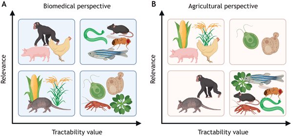
In their Commentary, Steadman, Vela and Burggren highlight the need to move away from traditional model animals in research on epigenetic inheritance, instead focussing on aligning model choice with the specific research question of interest.
Chimps’ wrists move the same as other apes’, despite different walking style
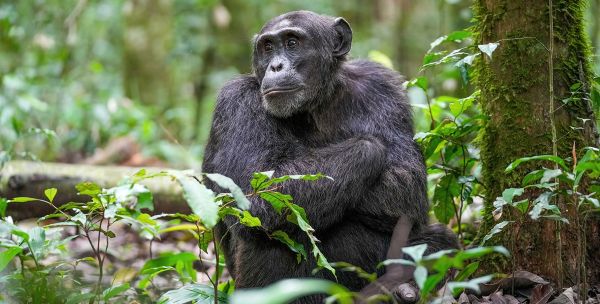
Walking on your knuckles is a weird way to get around, but it's normal for chimps and gorillas. Recently, Nathan Thompson and colleagues showed that although the hand posture is odd, the muscle contractions required are quite similar to a monkey's, which walk on their palms.














 English (US) ·
  French (CA) ·