

 1 month ago
74
1 month ago
74
PROTECT YOUR DNA WITH QUANTUM TECHNOLOGY
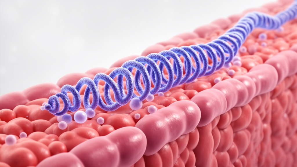
Orgo-Life the new way to the future Advertising by AdpathwayA new method reported in Nature Cell Biology is offering scientists an unusually detailed view of how chemical modifications are distributed across DNA and RNA inside tissues. The technique, called DREAMS, is designed to illuminate spatial landscapes of genetic and epigenetic information rather than treating a tissue as a uniform collection of cells. In practical terms, this means researchers can begin to ask not only which genes are chemically modified, but also where those modifications occur, which cells contain them, and how their locations relate to tissue structure. The study by Peng, Xue, Zhang and colleagues arrives at a moment when biology is moving beyond the simple reading of genetic sequences toward a more complete description of how cells regulate, edit and interpret molecular information. DNA and RNA modifications are central to that effort because they can influence gene activity, RNA stability, translation and cellular identity without changing the underlying genetic code.
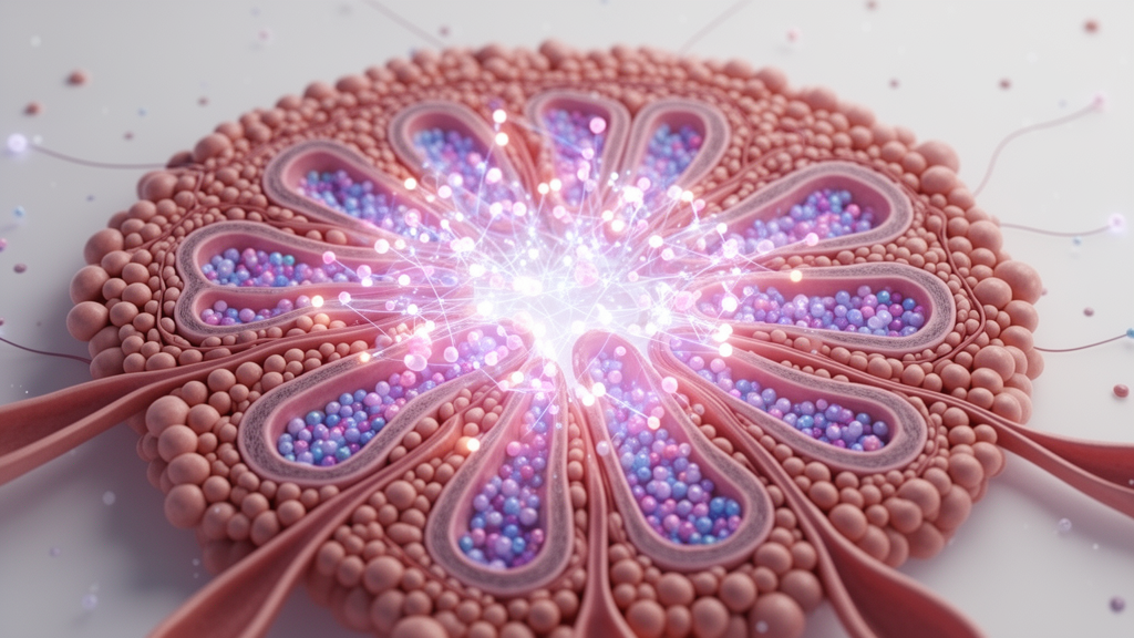
The significance of DREAMS lies in its attempt to examine DNA and RNA modification patterns within their native spatial context. Conventional molecular profiling methods often require tissues to be dissociated into individual cells or homogenized into a mixture. Those approaches can reveal the molecular characteristics of cell populations, but they may erase the physical relationships that make tissues function. A neuron next to a blood vessel, an immune cell positioned at the edge of a tumor, or a stem cell embedded within a specialized niche may behave differently from an otherwise identical cell in another location. By preserving spatial information, a technology such as DREAMS can connect molecular chemistry with anatomy. It has the potential to show whether a modification is concentrated in a particular cellular compartment, aligned with a structural boundary, or associated with communication between neighboring cells.
DNA modifications are among the most extensively studied chemical marks in biology. The best known is DNA methylation, in which a methyl group is added to cytosine bases, often at sites where cytosine is followed by guanine. Depending on genomic location and cellular context, methylation can contribute to gene silencing, genome stability and the maintenance of cell identity. It can also change during development, aging, disease and environmental stress. Yet DNA methylation is only one part of a broader regulatory system. Other chemical changes to DNA and its associated proteins can alter how tightly genetic material is packaged and how accessible it is to transcriptional machinery. Mapping these marks in space may reveal regulatory patterns that are invisible when molecular measurements are averaged across large numbers of cells.
RNA modifications add another layer of complexity. Messenger RNA, transfer RNA, ribosomal RNA and many regulatory RNA molecules can carry chemical marks that affect how they are processed and used. N6-methyladenosine, commonly known as m6A, is one of the best-known messenger RNA modifications and can influence RNA splicing, transport, stability and translation. Other modifications help specialized RNAs fold correctly or function efficiently during protein synthesis. These marks are dynamic: enzymes install them, remove them or interpret them, allowing cells to adjust gene expression after transcription has already occurred. A spatial map of RNA modifications could therefore provide information that a conventional gene-expression profile cannot. Two neighboring cells might produce similar amounts of an RNA while modifying it differently, potentially changing how long the molecule survives or how efficiently it is translated into protein.
DREAMS is particularly notable because it seeks to examine DNA and RNA modification landscapes together. In most experimental workflows, DNA epigenetics and RNA chemistry are studied through separate technologies, each optimized for a different type of molecule. Integrating them is technically difficult because DNA and RNA differ in structure, abundance, stability and chemical composition. DNA is relatively stable and present as a long-term archive, whereas many RNA molecules are short-lived and highly sensitive to degradation. A combined spatial approach must therefore preserve tissue architecture while distinguishing molecular species and retaining enough chemical information to identify modified bases. The resulting data can be viewed as a multilayered map: DNA modifications describe aspects of the regulatory state of the genome, while RNA modifications reflect how genetic messages are processed and deployed in individual regions of a tissue.
The technical challenge is not simply detecting whether a chemical mark exists. Researchers must also determine its position, its molecular identity and, ideally, its abundance. Chemical modifications can be subtle, and some may interfere with standard sequencing reactions in ways that are difficult to distinguish from technical noise. Spatial assays introduce additional complications, including limited molecular capture, uneven signal distribution and the need to align molecular measurements with microscopic images. A successful platform must combine chemical or enzymatic treatment with high-throughput detection and computational analysis. DREAMS appears to address this problem by linking modification-sensitive molecular readouts to spatially resolved measurements, allowing researchers to reconstruct where modified nucleic acids are located while maintaining the overall organization of the sample. That combination is what turns a molecular assay into a tissue-scale map.
The value of such maps becomes especially clear in complex biological systems. Development depends on precisely timed changes in gene regulation, and many of those changes involve DNA methylation, chromatin remodeling and RNA processing. In the nervous system, spatially restricted molecular programs help establish distinct neuronal populations and support communication between cells. In the immune system, local signals can alter the regulatory state of cells as they enter inflamed or infected tissues. Tumors are similarly organized into neighborhoods containing cancer cells, fibroblasts, blood vessels and immune populations, each influencing the others through chemical and physical signals. If DREAMS can distinguish modification patterns across these environments, it may help reveal why cells in the same tumor behave differently or why some regions resist treatment. The method could also provide a way to study how epigenetic and post-transcriptional regulation change as disease progresses.
One of the most important conceptual advances offered by spatial modification profiling is the possibility of connecting molecular marks with cellular states without relying entirely on predefined cell categories. Traditional analyses often classify cells according to gene-expression signatures and then compare modification levels between those groups. A spatial map can work in the opposite direction by revealing previously unrecognized molecular domains first. For example, a tissue may contain a narrow zone where DNA methylation changes gradually rather than abruptly, or a microenvironment where RNA modifications increase near a source of extracellular signaling. These patterns could point to transitions between cellular states, localized responses to stress or interactions that would be difficult to detect in dissociated samples. The approach may also help researchers distinguish cause from consequence by tracking whether a modification appears before or after a change in cell behavior.
As with any emerging technology, DREAMS will need to be evaluated across tissues, species and experimental conditions. Spatial measurements can be powerful, but they are not automatically complete. A technique may capture some classes of modifications more efficiently than others, and chemical processing can introduce biases related to sequence composition, transcript abundance or tissue preservation. Resolution is another key consideration: a signal assigned to a small region may still represent several nearby cells unless the method reaches single-cell or subcellular precision. Quantitative comparisons also require careful controls, calibration standards and validation with independent approaches. These limitations do not diminish the importance of the work; rather, they define the next stage of development. As protocols improve, spatial DNA and RNA modification maps could become more quantitative, more comprehensive and easier to integrate with transcriptomic, proteomic and imaging data.
The broader promise of DREAMS is that it treats genetic information as a living, spatially organized system rather than a static sequence. DNA modifications help establish which parts of the genome are available for use, while RNA modifications influence what happens to the messages copied from that genome. Their effects are shaped by cellular location, developmental history and communication with neighboring cells. By bringing these layers into the same experimental view, the method may help explain how tissues maintain order, how diseased regions emerge and how molecular states respond to treatment. The study does not replace existing sequencing or imaging technologies; instead, it adds a missing dimension to them. For researchers seeking to understand biology at the scale where cells actually operate, that dimension could prove decisive: not merely what molecular marks are present, but where they are, how they coexist and why their geography matters.
Subject of Research: Spatial mapping of DNA and RNA modification landscapes in tissues using the DREAMS technology.
Article Title: DREAMS illuminates spatial DNA and RNA modification landscapes.
Article References:
Peng, Y., Xue, L., Zhang, Y. et al. DREAMS illuminates spatial DNA and RNA modification landscapes.
Nat Cell Biol (2026). https://doi.org/10.1038/s41556-026-02038-z
Image Credits: AI Generated
DOI: https://doi.org/10.1038/s41556-026-02038-z
Keywords: DREAMS, spatial biology, DNA modifications, RNA modifications, epigenetics, RNA epitranscriptomics, DNA methylation, tissue mapping, molecular imaging, gene regulation
Tags: advanced methods for spatial genomicscellular heterogeneity in gene expressionchemical modifications in genetic regulationDNA methylation and histone modificationsepigenetic landscape visualizationmolecular profiling of tissuesRNA modification localizationspatial DNA and RNA modification mappingspatial epigenetics techniquesSpatial transcriptomicstissue architecture and gene regulationtissue-specific epigenetic landscapes


















 English (US) ·
  French (CA) ·